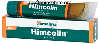
Himcolin dosages: 30 gm
Himcolin packs: 1 tubes, 2 tubes

Himcolin 30 gm buy
Occipitocervical fusion: indications most effective erectile dysfunction pills himcolin 30 gm cheap with mastercard, approach erectile dysfunction drugs associated with increased melanoma risk buy generic himcolin 30 gm, and long-term results in 13 sufferers. Occipito-cervicothoracic backbone fusion in a affected person with occipito-cervical dislocation and survival. Traumatic posterior atlantooccipital dislocation with Jefferson fracture and fracture-dislocation of C6�C7: a case report with survival. Posterior atlanto-occipital dislocation and concomitant discoligamentous C3-C4 instability with survival. Anterior C1�C2 screw fixation and bony fusion via an anterior retropharyngeal approach. Atlanto-axial arthrodesis by anterior retropharyngeal intermaxillo-hyoidal strategy [in French]. Salvage anterior C1C2 screw fixation and arthrodesis by way of the lateral method in a patient with a symptomatic pseudoarthrosis. Biomechanical evaluation of transoral plate fixation for atlantoaxial instability. Spine 2002;27:219�220 227 24 Craniocervical Disruption: Injuries of the Occiput�C1�C2 Region 123. One-stage posterior decompression and fusion using a Luque rod for occipito-cervical instability and neural compression. Fusion of the craniocervical transition with "CerviFix" after survived atlanto-occipital dislocation [in German]. Cervico-occipital fusion for congenital and posttraumatic anomalies of the atlas and axis. Luxation traumatique occipitoatloidienne: interet de nouveaux signes radiologiques (a propos de deux cas). Occipitocervical fusion with posterior plate and screw instrumentation: a long-term follow-up research. Occipitocervical arthrodesis using contoured plate fixation: an early report on a flexible fixation technique. An anatomic examine of the thickness of the occipital bone: implications for occipitocervical instrumentation. Biomechanical analysis of a brand new modular rod�screw implant system for posterior instrumentation of the occipito-cervical spine: in-vitro comparison with two established implant techniques. A biomechanical analysis of occipitocervical instrumentation: screw in contrast with wire fixation. Biomechanical evaluation of 5 totally different occipito-atlanto-axial fixation strategies. Clinical experience with rigid occipitocervical fusion in the administration of traumatic higher cervical spinal instability. Images in emergency medicine: long-term survival following full medulla/cervical spinal twine transection. Chiari I malformation related to atlanto-axial dislocation: specializing in the anterior cervicomedullary compression. Case reviews: Atlantooccipital and atlantoaxial traumatic dislocation in a child who survived. Irreducible anterior atlantoaxial dislocation: one-stage treatment with a transoral atlantoaxial discount plate fixation and fusion. Cranio-cervical stabilization of traumatic atlanto-occipital dislocation with minimal resultant neurological deficit. Novel therapy of basilar invagination ensuing from an untreated C-1 fracture associated with transverse ligament avulsion. Atlantal lateral mass screws for posterior spinal reconstruction: technical observe and case sequence. Avulsion transverse ligament accidents in children: successful treament with nonoperative management. Delayed decompression of persistent C1C2 subluxation in a pediatric patient with tetraplegia-is restoration possible Coronally oriented vertical fracture of the axis body: surgical therapy of a uncommon situation. A review of current options and early evaluation of rigid inside fixation techniques. Lindsey and Zbigniew Gugala the axis is a unique second vertebra that performs a important role in upper cervical spine movement and stability. The specialised anatomy and kinematics of the axis (in combination with the atlas) topics it to biomechanical hundreds that could be diverse and appreciable. These hundreds, when excessive, can lead to a selection of ligamentous and bony accidents, among which odontoid fractures are the most typical. Fractures of the odontoid can account for as a lot as 20% of all cervical backbone injuries. This classification system has been universally accepted and used to direct treatment. Symptomatic administration of the fracture with exercise modification and with out external supportive gadgets Nonoperative treatment (active). It can encompass posterior cervical fixation and fusion (using transarticular screws or sublaminar wires) or anterior fixation (using odontoid screws) Materials and Methods Literature Search An exhaustive database search was performed to determine all original printed studies pertaining to the subject. The publication database search included the National Library of Medicine and Medline; the search concerned all human studies printed in English between January 1966 and November 2007. The bibliography listed in these papers was evaluated for the presence of additional articles to guarantee a thorough and complete literature evaluation. Evaluating Articles and Weighing Strength of Evidence the literature chosen for evaluation was categorized in accordance with the strength of evidence. All chosen articles had been assessed by two impartial reviewers and categorized in accordance with their degree of proof. The levels of evidence determined for the odontoid fracture articles chosen had been then linked to remedy recommendations in accordance with the standards of the American Medical Association. Objectives and Paper Selection Criteria the target of this systematic evaluation was to tackle the following 4 questions: 1. For the purpose of this review aged was outlined as higher or equal to 60 years of age. Results the computerized search of the National Library of Medicine and Medline yielded a total of 443 papers revealed between May 1965 and November 2007. There was no level I proof research on odontoid fractures among the many selected articles; subsequently the authors had been unable to establish follow requirements in this chapter. A total number of 35 papers pertaining to Question 1 (halo vest vs brace) were recognized following aforementioned standards. Anderson et al prospectively studied V Upper Cervical Injuries and Their Management 230. Lind et al5 also prospectively analyzed eighty three unstable cervical backbone damage patients treated with the halo vest. Although the halo vest provides higher stability, its reported union rate can differ from 469 to 88%. A whole variety of 14 papers pertaining to Question 2 (nonoperative vs operative) have been recognized in accordance with the standards of the American Medical Association. Among the bigger studies, nonoperative therapy constantly demonstrated a excessive incidence of nonunion, which ranged from 20 as much as one hundred pc.
Hogweed (Knotweed). Himcolin.
- What is Knotweed?
- Bronchitis; cough; lung diseases; skin diseases; decreasing sweating with tuberculosis; increasing urine; redness, swelling, and bleeding of the gums, mouth, and throat; and preventing or stopping bleeding.
- Dosing considerations for Knotweed.
- Are there safety concerns?
- How does Knotweed work?
Source: http://www.rxlist.com/script/main/art.asp?articlekey=96539
Himcolin 30 gm low cost
However erectile dysfunction commercial bob himcolin 30 gm buy, now greater than ever a trend exists towards adopting new concepts solely within the presence of sound proof erectile dysfunction hypertension medications himcolin 30 gm online. This development is a product not only of the need to preserve a more scientific method on behalf of the surgeon, but can also be because of extra stringent ranges of fiscal accountability enforced by health care purchasers. The answers that we search, nonetheless, are sometimes hidden by the lack of adequately powered studies,1 a symptom of each the idiosyncrasies of spinal trauma and the issue in expressing the impression of an intervention on a person. Even in large studies where planning and execution appear properly conceived results may be unclear and conclusions overstated with an consequence where controversy remains the rule somewhat than the exception regardless of hundreds of thousands of dollars spent. This strategy permits for pooling of patient information to address the hitherto chronically underpowered spine literature. The profitable growth of national arthroplasty registers in Scandinavia has shown how powerful large multicenter databases are at recognizing trends, enabling well being care planning, and establishing acceptable standards. It publishes yearly statistical reports obtainable to the basic public in modified form. It is the biggest of its type with knowledge on almost 24,000 patients included in the most recent report. Continued improvement of disease-specific consequence measures will enhance the responsiveness and generalizability of databases to particular interventions. As the momentum builds for contribution to these giant databases, the number of mutually acceptable scoring methods will turn out to be paramount. These demographics underscore the necessity to contain highly populated nations in multinational databases to acquire a really global perspective. As has already been alluded to , the relevance of recent applied sciences for the majority of the two. An aspect, though much less glamorous in appeal, that is still basic to the important thing concern is decreasing the variety of individuals with disabilities and the severity of their disability. The urban inhabitants rose in China from 178 million in 1978 to 524 million in 2003,14 and in India from one hundred sixty million in 1981 to 285 million in 2001. Injury statistics are even additional magnified by, among different things, inadequate seatbelt compliance, more outstanding than may in any other case be predicted because of an absence of well being and security laws and failure to regulate such legislation the place it exists. It is vital that creating nations study from the successes and failures of almost 150 years of health and security legislation in established industrial nations. This might require continued pressure and monitoring where potential from worldwide health and labor organizations. Projects such as the China Seat Belt Intervention Study being sponsored by the George Institute for International Health in Guangzhou26 will help decide the feasibility of visitors laws enforcement and inhabitants education. Encouraging the development of spinal harm databases no less than in the giant urban areas of creating international locations should be thought of a priority. These would be the building blocks that may enable future variations of this chapter to have a truly world epidemiological part. Epidemiology of Spinal Column Injury Spinal column injury encompasses scientific diagnoses starting from gentle tissue cervical spine accidents, by way of low-energy insufficiency fractures, high-energy fracture dislocations, and finally full cord harm. The obvious explosion in incidence of whiplash-associated disorders coincides with elements together with elevated seatbelt use, visitors congestion, and an increasingly litigious tradition within the West. The incidence and prevalence have additionally contributed to the development of vertebral augmentation techniques. Yet long-term morbidity from these fractures is low, with up to two thirds being recognized only by the way on unrelated radiographs. The vast majority of low-energy, symptomatic fractures are self-limiting and respond well to nonoperative measures. With the appearance of fit, cell, adventure-seeking septa- and octogenarians a greater diploma of surgical cases could be expected. Presently although, epidemiological information like these are likely to be fragmented and geographically isolated in nature. Tremendous progress has been made preventing injuries in sports activities such as diving40�42 and rugby43 as a end result of instructional harm prevention packages. However, in new thrill-seeking sports such as snowboarding44,forty five and mountain biking46 there has been a rising variety of spinal injuries, and future studies should determine the epidemiology of these injuries and the effect of protective equipment in order that harm prevention programs can be targeted. Fall-induced injuries represent a major well being drawback highlighting a necessity for fall prevention measures and proactive therapies for osteoporosis. Unsurprisingly, the variety of patients discharged from hospital on a ventilator has increased from 2. Similar tendencies in survival are being reported in different industrialized international locations corresponding to Denmark and the United States. The sequelae that develop following injury are also in contrast with nonobese sufferers (32% vs 16%, p zero. Two questions on this field that stay difficult to answer involve imaging of the cervical spine (1) in the patient with minor trauma, and (2) in the unconscious polytrauma affected person. The Canadian C Spine Rules, a prospective research that developed tips for when to get hold of plain radiographs within the alert trauma affected person, has addressed the previous drawback. Despite these quantum leaps in imaging, scientific acumen remains elementary to figuring out pretest probability and thus attaining the highest diagnostic accuracy. In the acute trauma phase one must decide whether or not the danger of delaying treatment to get hold of further imaging is outweighed by the benefit of further data. This quandary is illustrated by the affected person with an incomplete neurological deficit ensuing from a cervical side harm requiring discount and the chance of a damaged disk inflicting further neurological insult on manipulation. Changes within the Management of Spine and Spinal Cord Injuries Spine Trauma Care Systems Trauma care in industrialized nations has developed from local to regional care techniques. Such applications ship treatment pathways that span the continuum from prevention, through acute care, and to reintegration into the neighborhood. For the backbone trauma community regionalization has tremendous benefits, including enhancing standardized care, accessing patients for clinical trials, creating benchmarks for national spinal trauma outcomes facilitating population-based studies by way of registries, and having the ability to intervene in a timely trend with new restore and regeneration interventions ought to these become available. Spine trauma registries have become extremely refined and might now provide data essential to understanding and bettering patient care. Statistics are slowly changing into obtainable detailing the variety of prehospital deaths, survival following hospital discharge. Absence of organized prehospital care in some growing countries is in sharp distinction to advanced techniques in some industrialized countries where a skilled physician can be rapidly positioned on the scene of the damage enabling optimized acute management. The advantages of prehospital care have been documented in the spine trauma population. Advances in prehospital screening have lowered the incidence of misdiagnosis within the field from 19 to 5%,36,forty three leading to less neurological deterioration. Further analysis is required to determine which patients need to be immobilized and how they should be immobilized. Diagnostic imaging has enabled accurate spinal wire and column visualization, not solely enhancing preoperative planning for implant placement but in addition directing the choice of surgical method to enable enough decompression. As a results of advances in biomaterial properties and new surgical strategies restoration of spinal column alignment has turn out to be comparatively easy in contrast with the challenges confronted by previous generations of spinal surgeons. Examples of this evolution could be present in anterior cervical locking plates and posterior segmental inflexible fixation methods, 34 which arguably render the halo vest somewhat out of date. Halo vest immobilization remains a gold normal for discount and stabilization of odontoid fractures; however, anterior odontoid fixation has gained widespread reputation.

Himcolin 30 gm order with visa
These strategies might not properly cut back fractures or tackle mechanical instability erectile dysfunction for young males himcolin 30 gm purchase amex. This practice might scale back the number of complications such as implant failure and decrease the necessity for greater morbidity anterior approaches erectile dysfunction homeopathic proven 30 gm himcolin. Preliminary observe on the treatment of vertebral angioma by percutaneous acrylic vertebroplasty [in French]. Long-term observations of vertebral osteoporotic fractures treated by percutaneous vertebroplasty. Reinforcement of thoracolumbar burst fractures with calcium phosphate cement: a biomechanical examine. No severe process related complications have been reported on this limited variety of collection. Most of the articles report some nonsymptomatic cement leakage, normally anterior, lateral or intradiskal. Potentially these are methods that will trigger harm to the patient in inexperienced arms. From the available, very 503 fifty one Vertebroplasty Techniques for Stabilization of Thoracolumbar Spine Traumatic Fractures to achieve good injectability. Balloon vertebroplasty with calcium phosphate cement augmentation for direct restoration of traumatic thoracolumbar vertebral fractures. Percutaneous transpedicular vertebroplasty with calcium phosphate cement within the therapy of osteoporotic vertebral compression and burst fractures. Kyphoplasty-augmented short-segment pedicle screw fixation of traumatic lumbar burst fractures: preliminary medical experience and literature evaluate. Treatment of thoracolumbar fractures with vertebroplasty together with posterior instrumentation. Direct discount of thoracolumbar burst fractures by means of balloon kyphoplasty with calcium phosphate and stabilization with pedicle-screw instrumentation and fusion. Prospective research of standalone balloon kyphoplasty with calcium phosphate cement augmentation in traumatic fractures. The treatment of acute thoracolumbar burst fractures with transpedicular intracorporeal hydroxyapatite grafting following oblique reduction and pedicle screw fixation: a potential study. Balloon vertebroplasty in combination with pedicle screw instrumentation: a novel technique to treat thoracic and lumbar burst fractures. Spine 2005; 30:E73�E79 fifty two Minimally Invasive Posterior Stabilization Techniques in Trauma Neel Anand, Eli M. Dekutoski Posterior minimally invasive strategies are novel techniques used within the treatment of spinal trauma. Theoretically they might be associated with lowered morbidity, blood loss, and muscle harm compared with traditional open methodologies. As enabling technologies develop, the applying of minimally invasive, posterior spinal stabilization techniques in the administration of spinal trauma continues to increase. However, the present stage of proof to support the utilization of these techniques compared with open techniques remains weak. Several small case collection have demonstrated feasibility and safety of these methods. However, evidence to help superiority and therefore suggestions for widespread adoption of these strategies compared with current open methods is missing. The epidemiology, distinctive clinical and diagnostic features as well as the preliminary administration of accidents pertinent to this chapter are discussed within the particular person chapters specific to every injury. Verlaan et al reported a median blood loss larger than 1000 mL for fixation of thoracolumbar trauma. Surgical Options Pedicle screw fixation has turn into the mainstay technique for repairing unstable thoracolumbar spine fractures, particularly with regard to providing stability within the setting of an incompetent posterior ligamentous complicated. Good outcomes with pedicle screw fixation for thoracolumbar fractures have been reported; however, a higher failure rate has been described in cases of anterior column insufficiency. Prior to performing the process, the affected person ought to have acceptable workup and imaging. This methodology is used to insert Jamshidi needles on one aspect and then the other. Subsequently, guide wires are launched down the Jamshidi needle underneath lateral fluoroscopic steering and the Jamshidi needles are eliminated. We prefer that the surgeon maintain the information wire with the left hand while holding the handle of the working cannula. A drill is used to drill a channel into the vertebral physique or a curette is used to tamp the vertebral body. Afterward the balloon is deflated and polymethylmethacrylate cement is introduced into the cavity created by the balloon. Should cement be seen exiting the cannula posteriorly, the technique is stopped to allow the cement to harden. By waiting a couple of moments, the surgeon permits the cement posteriorly to stiffen; thus any cement launched further is much less likely to advance in the same direction. Cement must be introduced till it fills the void created by the balloon and interdigitates with the bony fragments. We choose to see the vertebral end plate top restored; however, nice care ought to be taken to avoid cement exiting the vertebral body either laterally or posteriorly. For placement of percutaneous pedicle screws, serial dilators are positioned over the guide wires. Care must be taken to make positive that pores and skin incisions are giant sufficient to accommodate the dilators. Subsequently, a cannulated tap is placed over the guide wire and used to faucet each pedicle. Once the pedicle screw is superior past the bottom of the pedicle, the guide wire may be eliminated. Additionally, extenders ought to have their slots at roughly the identical heights. Once the rod is inserted into the primary pedicle screw extender, a tester is use to make sure that the rod is definitely within the extender and never lateral to it. Lateral fluoroscopy is used to confirm acceptable depth and extender orientation. Once seated, a suction trephine is used to suck out any delicate tissue contained in the extenders, and a prime locking nut is placed within the caudal screw and tightened. The surgeon has the option of applying compression across the screws previous to tightening down all of the nuts. The pores and skin incisions are prolonged between the screw heads, the fascia is split, and the sides are then uncovered, irrigated, and decorticated. A bone graft extender or osteobiological merchandise and native bone graft are positioned. For proof concerning the utilization of cement augmentation for traumatic spinal fractures, please see Chapter 51. Image steerage has been used with percutaneous screw placement and may have an rising position in the future.

30 gm himcolin discount with visa
The external indirect muscular tissues serve to present contralateral axial rotation of the trunk can you get erectile dysfunction young age purchase himcolin 30 gm free shipping, whereas the inner obliques prostaglandin injections erectile dysfunction discount himcolin 30 gm on line, together with the transversus abdominis, oppose this motion by way of an ipsilateral axial rotation of the trunk. However, pure flexion of the lumbar backbone is usually the obligation of the rectus abdominis. Moving extra inferiorly into the lumbosacral region, the quadratus lumborum and iliopsoas muscular tissues are responsible for ipsilateral truncal bending. The cervical spine and thoracolumbar spine are essentially the most mobile areas of the spinal axis. Range of movement in the cervical backbone is necessary for head motion and direction of gaze. In the frontal aircraft, the cervical spine undergoes 35 levels of lateral flexion when an angle is shaped between a line extending laterally from the clavicle and another line extending downward from the lateral angle of the attention. In the sagittal aircraft, the cervical backbone can normally flex sixty five levels, bringing the chin towards the chest, and extend 40 levels. The normal diploma of axial rotation with respect to the sagittal plane is 50 levels bilaterally. In the thoracolumbar spine, contributions to vary of motion may be divided between the thoracic and lumbar areas. This produces a total thoracolumbar ahead flexion of eighty five degrees utilizing the larger trochanter of the hip as the fulcrum of motion and the acromion as the dynamic point of reference. Using the identical landmarks, thoracolumbar extension is the sum of 25 levels of extension within the thoracic spine and 35 degrees of extension in the lumbar backbone. This motion is equally divided between thoracic and lumbar motion, which is measured using the S1 vertebral body as the fulcrum and the L1 vertebra and the C7 vertebra because the dynamic factors of reference for lumbar and thoracic lateral flexion, respectively. Axial rotation primarily happens within the thoracic backbone and is split into 35 degrees within the thoracic spine and 5 degrees in the lumbar spine. Total movement of the spine during any particular motion may be estimated by the addition of actions of every spinal region. For instance, in a standard affected person, whole forward flexion cervical (65 degrees) thoracic (35 degrees) lumbar A. Not surprisingly, the cervical spine is essentially the most flexible in all planes of motion. Vasculature the blood provide to the cervical spinal column is normally derived from branches of the subclavian artery distal to the origin of the widespread carotid arteries. The first three cervical vertebrae most frequently receive their blood provide from the vertebral artery, whereas the fourth through sixth cervical vertebrae are typically provided by the ascending cervical artery, which arises from the thyrocervical trunk. The seventh cervical vertebra is provided by the deep cervical artery, which arises from the costocervical trunk. This artery, in turn, divides into three elements: the anterior and posterior spinal canal branches and the radicular artery. The anterior and posterior spinal canal branches provide the vertebral buildings and ligaments, whereas the radicular artery continues to journey along the nerve root and eventually enters the subarachnoid space and bifurcates into an anterior and posterior department. Both of these vessels sometimes arise from the vertebral arteries and descend the length of the spinal twine. In some circumstances, the posterior spinal arteries may also arise from the posterior inferior cerebellar artery. The radiculomedullary arteries provide collateral move between the anterior and posterior spinal arteries and sure radicular arterial branches. These collaterals are essential in sustaining Neuroanatomy the neuroanatomy of the spinal cord is constant all through its size from the foramen magnum to the lumbar backbone the place it usually terminates in the area of the primary lumbar vertebra. Grossly, the spinal wire steadily turns into smaller in diameter as it descends because of progressively much less afferent and efferent axons because the spinal nerves branch off by way of the neural foraminae. In these areas the spinal wire turns into larger in cross part as the numerous motor and sensory neurons together with their related interneurons which kind the brachial and lumbar plexus take part in rich synaptic networks. The preponderance of anterior horn cells additionally contributes to the increased spinal wire diameter seen in these regions. Histologically, the periphery of the spinal cord is composed of myelinated white matter, whereas the central "H-shaped" space consists of unmyelinated grey matter. The posterior funiculus of the spinal wire incorporates the dorsal column medial lemniscus pathway, which transmits sensory data including fine touch, vibration, and proprioception. Degeneration or a lesion in this area will predictably compromise these sensory discriminations and severely impair joint and vibratory sense distal to the lesion. The spinothalamic tract is split into an anterior portion, which transmits touch alerts, and a lateral portion, which transmits alerts for pain and temperature. Impulses inside these tracts are routed from the periphery to the thalamus for central processing and integration. The other primary perform of the spinal wire is to present distal motor innervation. The anterior funiculus and a portion of the lateral funiculus comprise the pyramidal and extrapyramidal motor tracts from their origin in the mind via the twine. Both voluntary and involuntary motor impulses are transmitted by way of these tracts. The spinal cord additionally relays motor and sensory data of the autonomic nervous system to control visceral perform. Within the gray matter core of the spinal wire as seen on cross-section lie anterior and posterior horns. The axons that have their cell our bodies in the anterior horn are lower motor neurons which are synapsing with the regulatory higher motor neurons. These axons exit from the anterolateral facet of the spinal twine segment because the ventral roots and merge with the dorsal root to make up the spinal nerve, which then exits the spinal column through the neural foramen. In the posterior horn is the tract of Lissauer, which is the first synapse of the fibers coming from the neuron within the dorsal root ganglion to transmit their signals through the secondary axons to the thalamus for central processing. This tract, together with the posterior funiculus, receives its blood supply from the posterior spinal arteries. The implications of surgical intervention in the backbone have to be carefully thought of when planning spinal procedures, and the intricate stability of the spinal column must be preserved to whatever extent possible to present maximum perform and stop or decrease pain. Only by remaining a vigilant student of the origin, structure, and function of the backbone can one provide the absolute best take care of the spinal patient. Prenatal growth of the traditional human vertebral corpora in several segments of the spine. Congenital abnormalities of the urogenital tract in association with congenital vertebral malformations. The contribution made by a single somite to the vertebral column: experimental evidence in help of resegmentation using the chick-quail chimaera mannequin. Philadelphia: Lippincott, Williams and Wilkins; 2001:93 Summary Study of the embryology and anatomy of the spine is of nice importance for practitioners caring for spine sufferers. The advanced three-dimensional structure of the backbone also can make analysis and treatment of spinal disorders quite difficult. Atlantoaxial rotatory subluxation in sufferers with Marfan syndrome: a report of three circumstances. Biomechanics and biochemistry of the intervertebral disks: the necessity for correlation research. J Bone Joint Surg Br 1974;fifty six:225�235 7 Emergency Room Evaluation of the Spinal Injury Patient Including Assessment of Spinal Shock Bizhan Aarabi and Yasutsugu Yukawa Spinal Shock and Digital Rectal Examination in the Emergency Room How can we grade the anatomical and useful integrity of the spinal wire after an injury The presence of spinal shock can be historically related to a poor prognosis.

Himcolin 30 gm generic overnight delivery
Surprisingly erectile dysfunction boyfriend buy discount himcolin 30 gm online, nonetheless impotence cream himcolin 30 gm order without prescription, few methods have been proposed to quantify this harm feature. In a cadaveric research, Isomi et al36 included measurements of the anterior and posterior vertebral body heights as part of their assessment of experimental thoracolumbar burst fractures. In the present systematic review, a myriad of articles and chapters allege the importance of so-called important thresholds for vertebral body height loss. Because reproducibility has yet to be assessed, this work provides only degree V proof that the prescribed measurement methodology is a reasonable current normal. Vertebral Body Translation Vertebral body translation is a typical radiographic function following thoracolumbar trauma. It signifies that a substantial quantity of force has been delivered to the spine, often leading to ligamentous disruption. In the present systematic evaluation, just one examine detailed a technique of measuring translation of thoracolumbar fractures. The interobserver or interobserver reliability of this measure was not reported, nor has it been subsequently assessed. Sacrum the sacrum is the transition zone between the cellular backbone and the pelvis, and in most centers, sacral fractures are handled by orthopedic traumatologists. Thus the large majority of the literature and terminology concerning description and measurement of those injuries lies throughout the orthopedic trauma literature. Data might be derived primarily from the recent evaluation by Kuklo et al,6 who published recommendations on behalf of the Spine Trauma Study Group. However, the authors admittedly recognize the lack of standardization and broad variability by which this measurement is made within the literature. These information provide solely level V evidence that the Spinal Canal Compromise Spinal canal compromise is a frequent radiographic finding after thoracolumbar trauma. The potential relationship between posttraumatic spinal canal encroachment and the presence or severity of neurological deficit has been studied extensively by way of both stenosis predisposing to harm and traumatic canal deformation. These include the sagittal canal diameter, the transverse canal diameter, the sagittal:transverse ratio, total canal crosssectional area, and proportion canal occlusion. Furthermore, the inter- and intraobserver reliability of these measures has not been assessed. In a retrospective examine, Vaccaro et al33 found that the propensity for neurological injury correlated more highly with the sagittal:transverse ratio than to the absolute space of the spinal canal. Rasmussen et al34 in contrast a computerdigitized methodology to a manually calculated methodology of assessing the cross-sectional space of the spinal canal, finding that the previous correlated well with the latter. Hashimoto et al 35 calculated the ratio of the canal space at the injured segment in relation to uninjured segments. This group claimed to discover crucial values above which a neurological deficit was extra probably. Summary Achieving standardization among a various population of surgeons and researchers is difficult, particularly when considering an entity as protean as backbone trauma. Nomenclature and measurement schemata have developed in parallel, drawing from the insights of a diverse range of experienced observers, and mirror the progress of know-how out there to diagnose and treat spinal trauma. Fundamental to any clinically relevant classification system, the standardization of terminology as related to the definition of harm traits is important to figuring out optimum therapy choices. Ideally a common canon of measurement strategies could be utilized to every area of the spinal column, and the lexicon of spinal trauma can be fixed regardless of surgical discipline. Although heterogeneity nonetheless exists, the Spine Trauma Study Group has been working towards a objective of standardization of these parameters. Only by way of prospective evaluation and objective validation will the group be able to decide the ultimate utility of these or another evaluation methods. Measurement methods for lower cervical backbone accidents: consensus statement of the Spine Trauma Study Group. Measurement methods for upper cervical spine injuries: consensus statement of the Spine Trauma Study Group. Radiographic measurement parameters in thoracolumbar fractures: a systematic evaluate and consensus statement of the Spine Trauma Study Group. Jefferson fractures: the role of magnification artifact in assessing transverse ligament integrity. Intra- and inter-rater reliability of the anterior atlantodental interval measurement from typical lateral view flexion/extension radiographs. Fractures of the ring of the axis: a classification based mostly on the analysis of 131 circumstances. Use of computed tomography to predict failure of nonoperative therapy of unilateral aspect fractures of the cervical backbone. The optimal radiologic methodology for assessing spinal canal compromise and cord compression in patients with cervical spinal twine damage, I: An evidence-based evaluation of the published literature. Interobserver and intraobserver reliability of maximum canal compromise and spinal twine compression for evaluation of acute traumatic cervical spinal cord damage. Cobb method or Harrison posterior tangent method: which to choose for lateral cervical radiographic analysis. Measurement of lumbar lordosis: analysis of intraobserver, interobserver, and technique variability. Comparison of computerized tomography parameters of the cervical backbone in normal control subjects and spinal cord-injured patients. Reciprocal angulation of vertebral bodies in a sagittal aircraft: method to references for the evaluation of kyphosis and lordosis. The significance of thoracolumbar spinal canal measurement in spinal twine damage sufferers. Reduced transverse spinal space secondary to burst fractures: is there a relationship to neurologic injury Relationship between traumatic spinal canal stenosis and neurologic deficits in thoracolumbar burst fractures. Radiographic parameters for evaluating the neurological spaces in experimental thoracolumbar burst fractures. Vaccaro the determination of spinal stability is probably considered one of the most important duties confronting spinal specialists as its presence considerably affects the therapy strategy of accidents to the spine. This dedication, nevertheless, stays challenging and continues to evolve, for significant ambiguity exists in its definition despite widespread use of the term. The concept was first launched in the Watson-Jones classification of spinal fractures in 1931 after which verified by Nicoll in 1949. The trendy idea of mechanical stability of the spinal column was described by White and Panjabi. Denis also concluded that a backbone that would withstand normal physiological stresses with out progressive deformity or neurological abnormalities, or each, was thought of stable. The challenge lies in the classes that make up the grey zone corresponding to burst fractures, disk disruptions, and certain flexion�distraction accidents. At current, final determination of spinal stability requires consideration of many different elements taken on a case-by-case basis. By far the most typical occipital cervical dislocation is a longitudinal separation, though posterior and anterior dislocations could occur. The relationship of the dens to the basion and the Power ratio are useful for diagnosis of ligamentous disruption and instability. In a standard cervical spine, the space from the tip of the odontoid to the basion is 4 to 5 mm in adults and as much as 10 mm in kids.

Buy himcolin 30 gm without a prescription
Vitamin K also functions in bone calcification; g-carboxylates glutamate residues in osteocalcin impotence sentence examples himcolin 30 gm low price. Vitamin K deficiency is rare impotence news himcolin 30 gm buy mastercard, however can be caused by the use of broad-spectrum antibiotics, which destroy colonic bacterial synthesis of the vitamin. Calcium features in bone formation, nerve conduction, muscle contraction, blood clotting, and cell signaling hypocalcemia produces tetany. Sodium features in acid-base steadiness, osmotic stress, muscle and nerve excitability, active transport, and membrane potential; deficiency produces abnormalities in mental status and convulsions. Potassium functions in acid-base steadiness, osmotic stress, muscle and nerve excitability, and insulin secretion; deficiency produces muscle weakness and polyuria. Phosphate features in bone formation, nucleotide structure, metabolic intermediates, metabolic regulation, vitamin operate, and acid-base balance; deficiency produces muscle weak spot, rhabdomyolysis, and hemolytic anemia. Chloride functions in acid-base stability, osmotic strain, and nerve and muscle excitability; deficiency signs are undefined. Sources of calcium include dairy merchandise, leafy green vegetables, legumes, nuts, and whole grains. Parathyroid hormone will increase reabsorption in the early distal tubule of the kidneys and mobilizes calcium from bone. Approximately 40% of calcium is certain to albumin; 13% is sure to phosphorus and citrates; and 47% circulates as free, ionized calcium, which is metabolically lively. Clinical findings related to hypomagnesemia and hypermagnesemia (see Table 4-3) D. Aldosterone: controls renal reabsorption (when present) and excretion (when absent) c. Inappropriate secretion of antidiuretic hormone: dilutional impact in plasma of excess water reabsorption from the accumulating tubules of the kidneys c. Sodium: acid-base stability, osmotic stress, muscle and nerve excitability, lively transport, and membrane potential Sodium controls water movement between extracellular and intracellular fluid compartments. Aldosterone: controls renal reabsorption (when absent) and excretion (when present) b. Zinc capabilities as a cofactor for metalloenzymes; deficiency produces poor wound therapeutic, dysgeusia, anosmia, and progress retardation. Copper features as a cofactor for metalloenzymes and in cytochrome oxidase; deficiency produces microcytic anemia, aortic aneurysm, and poor wound therapeutic. Selenium functions in antioxidant action as a component of glutathione peroxidase; deficiency produces muscle pain and weak spot. Ferritin, a soluble iron-protein advanced, is the storage type of iron in the intestinal mucosa, liver, spleen, and bone marrow. Hemochromatosis: cirrhosis of the liver, bronze pores and skin shade, diabetes mellitus, malabsorption, and coronary heart failure Transferrin: capabilities in iron transport Low iron stores: transferrin increased Iron shops excessive: transferrin decreased Iron poisoning (1) Common in kids (2) Causes hemorrhagic gastritis and liver necrosis b. Iron overload illnesses: hemochromatosis; hemosiderosis; sideroblastic anemia (due to pyridoxine deficiency, lead poisoning, alcoholism) (1) Sideroblastic anemias are related to extra iron accumulation in mitochondria resulting from difficulties in heme synthesis. Various illnesses (1) Alcoholism, rheumatoid arthritis, acute and continual inflammatory diseases, continual diarrhea b. Acrodermatitis enteropathica (1) Autosomal recessive illness associated with dermatitis, diarrhea, development retardation in youngsters, decreased spermatogenesis, and poor wound healing 5. Sources of copper embody shellfish, organ meats, poultry, cereal, fruits, and dried beans. Chromium is a component of glucose tolerance issue, which facilitates insulin motion through post-receptor results. Selenium is a element of glutathione peroxidase, which converts oxidized glutathione (see Chapter 6) into lowered glutathione within the pentose phosphate pathway. Deficiency of fluoride is primarily due to inadequate consumption of fluoridated water. Glutathione: antioxidant that neutralizes peroxide and peroxide free radicals Selenium: component of glutathione peroxidase Fluoride: structural component of hydroxyapatite in bone and enamel Fluorosis: chalky deposits on the enamel, calcification of ligaments, an increased threat for bone fractures Fluoride deficiency: dental caries H. A lower in free vitality for a biochemical reaction, or sequence of reactions, indicates its tendency to proceed. A reaction that requires a free power input must be coupled to one other reaction that releases no less than that a lot power. Metabolic pathways encompass a series of coupled reactions linked by frequent intermediates. In the absence of such processes, particular person reversible reactions finally attain equilibrium, and the flow of metabolites through a pathway ceases. For instance, a genetic defect or inhibitor that reduces manufacturing of B additionally decreases operation of the pathway from fuel! There are five frequent elements of metabolic pathways: response steps, regulated steps, distinctive characteristics, pathway interfaces, and scientific relevance. Acetyl CoA is utilized in fat synthesis, ldl cholesterol synthesis, ketone physique synthesis, and formation of acetylated molecules. Inner membrane: oxidative phosphorylation Acetyl CoA: product of fats and glucose oxidation Acetyl CoA: a focal point in metabolism Glycolysis, glycogenesis, glycogenolysis, pentose phosphate shunt, fatty acid synthesis, steroid synthesis. Many intermediates within one pathway are substrates for other pathways, providing a way for different pathways to interact. Pyruvate carboxylase, which varieties oxaloacetate by carboxylation of pyruvate, is allosterically activated by acetyl CoA. A deficiency of any of those vitamins negatively impacts operation of the cycle and impairs vitality production. Cycle intermediates additionally participate in artificial pathways leading to glucose, fatty acids, porphyrins, and amino acids (dashed arrows). Because all mitochondria within the zygote come from the ovum, these illnesses exhibit maternal inheritance, by which affected moms transmit the illness to all of their youngsters. Uncouplers short-circuit the proton gradient by transporting H� ions from the intermembrane area to the matrix, thereby abolishing the gradient. Pyruvate dehydrogenase is regulated by covalent modification with phosphorylation. Glycolysis interfaces with glycogen metabolism, the pentose phosphate pathway, the formation of amino sugars, triglyceride synthesis (by means of glycerol 3-phosphate), the production of lactate (a dead-end reaction), and transamination with alanine. Pyruvate dehydrogenase interfaces with different pathways such because the citric acid cycle or fats synthesis by way of its product, acetyl CoA. Deficiencies in any of the pyruvate dehydrogenase enzymes produce lactic acidosis. Phosphorylation of glucose to glucose 6-phosphate, the first regulated step in glycolysis, is irreversible and traps glucose inside the cell. Glucokinase, present within the liver and pancreatic b cells, is very lively only at excessive glucose concentrations (high Km) and rapidly phosphorylates massive amounts of glucose (high Vmax). Reversible conversion of fructose 1,6-bisphosphate to two 3-carbon intermediates by aldolase A b. The regulated steps in glycolysis are indicated by one-way arrows and boxed enzymes. Reversible conversion of 3-phosphoglycerate to 2-phosphoglycerate by phosphoglycerate mutase eight. This reaction happens in anaerobic glycolysis related to shock and extreme exercise.
Syndromes
- Do not sit or stand for long periods. Even moving your legs slightly helps keep the blood flowing.
- Chest x-ray or chest CT scan
- Return from surgery with a Foley catheter in your bladder
- There is pus or blood in your stools.
- Damage to the kidneys
- Laxative
- Placing a small tube called a stent into an artery to help hold it open
- Irregular, fast heart rhythms (arrhythmias)
- Sudden and urgent need to urinate (urinary urgency)
Himcolin 30 gm purchase mastercard
Similarly impotence guilt purchase himcolin 30 gm line, the idea of early decompression of compromised neural parts has remained a key tenet within the therapy of spinal cord damage doctor for erectile dysfunction in delhi 30 gm himcolin cheap overnight delivery. This concept is, nevertheless, solely relevant with some limitations for sufferers with sacral fractures due to the various elements recognized in the preceding part on remedy options. We identified a total of 260 sufferers in six studies discussing outcomes of affected person survival or useful restoration related to timing of intervention. In a retrospective examine using historic controls for sufferers with unstable pelvic ring fractures, Latenser et al discovered statistically decreased hospital stays, lowered blood transfusion necessities, and decreased disability when sufferers had been treated before 72 hours. As to timing of intervention having an influence on neurological outcomes, the evidence base is totally anecdotal. The authors observed tougher fracture reduction after seventy two hours following harm due to presumed "muscle contractions. The analysis and therapy of sacral fractures has changed dramatically with 448 the arrival of newer imaging methods permitting more accurate diagnosis and understanding of fracture patterns. Diagnosis of neurological damage has been improved in each the alert in addition to the obtunded patient with neurodiagnostic research. Treatment of sacral fractures continues to evolve with bettering constructs and implants. Treating surgeons now have at their disposal options starting from simple decompression to complete lumbopelvic fixation. The overall clinical image of the affected person is of paramount importance and will affect therapy accordingly. As diagnostic and therapy modalities continue to evolve, will in all probability be up to the surgeon to remain abreast of these adjustments and adapt accordingly. To date the general power of proof for any type of care of sacral fractures is weak, whereas the strength of treatment advice based mostly upon the timing of intervention is modest. Treatment advice is robust as to the need for surgical therapy of less displaced fractures with percutaneous fixation and possible neural factor decompression and open discount and inside fixation for unstable fractures (1 cm displacement and neurological deficit) utilizing some type of segmental low-profile lumbopelvic fixation. American Spinal Injury Association and International Medical Society of Paraplegia. Diagnosis and management of closed internal degloving injuries related to pelvic and acetabular fractures: the Morel-Lavall�e lesion. Honda signal and variants in sufferers suspected of getting a sacral insufficiency fracture. Sacral insufficiency fractures caudal to instrumented posterior lumbosacral arthrodesis. Radiographic measurement techniques for sacral fractures consensus statement of the Spine Trauma Study Group. Intraoperative somatosensory evoked potential monitoring throughout acute pelvic fracture surgical procedure. Lumbosacral nerve injury in fracture of the pelvis: a postmortem radiographic and patho-anatomical examine. Hemorrhage associated with pelvic fractures: causes, analysis, and emergent administration. Midline sagittal sacral fractures in anterior-posterior compression pelvic ring injuries. Failure of reduction with an external fixator within the management of injuries of the pelvic ring: long-term evaluation of one hundred ten sufferers. Sexual operate after major resections of the sacrum with bilateral or unilateral sacrifice of sacral nerves. Treatment of unstable pelvic fractures: use of a transiliac sacral rod for posterior lesions and an external fixator for anterior lesions. Long-term useful prognosis of posterior injuries in highenergy pelvic disruption. Assessment of pelvic ring stability after damage: indications for surgical stabilization. Radiographic analysis of sagittal airplane alignment and balance in standing volunteers and patients with low again ache matched for age, intercourse, and measurement: a potential controlled clinical examine. Functional consequence of open reduction and inner fixation for completely unstable pelvic ring fractures (type C): a report of 40 circumstances. Percutaneous stabilization of U-shaped sacral fractures utilizing iliosacral screws: approach and early outcomes. Triangular osteosynthesis and iliosacral screw fixation for unstable sacral fractures: a cadaveric and biomechanical evaluation beneath cyclic hundreds. Anatomic and radiographic concerns for placement of transiliac screws in lumbopelvic fixations. This chapter offers a scientific, evidencebased-medicine evaluation of the literature in regards to the fundamental aspects of this entity, particularly, the following questions might be analyzed and answered by way of this review process: 1. The regional investigation of noradrenaline spillover release demonstrated important increases only below the lesion stage. Intact carotid and aortic baroreceptors are activated by the hypertension and set off the brainstem reflexes to lower the blood stress. It could also be only a relative slowing of the center; generally it could not drop as little as 60 beats per minute, however it can be insufficient to compensate for the vasoconstriction. The second compensatory mechanism is a rise within the inhibitory outflow from vasomotor centers above the lesion to dilate the splanchnic bed to accommodate the excessive amount of blood resulting from the increased peripheral resistance. However, the inhibitory impulses are unable to move under the injury, and dilation of vascular beds is proscribed to above the harm degree, leading to profuse sweating and skin flushing. Intact sensory nerves under the damage degree transmit noxious or non-noxious impulses that probably induce a widespread activation of the sympathetic nervous system. Cardiac arrhythmias, atrial fibrillations, and untimely ventricular contractions have been reported in the course of the persistent phase. People in cost of these patients should pay consideration to the variability of the described symptoms that could be minimal and even absent. People with low verbal communication ability can have issue expressing their signs. Elevated Blood Pressure the primary symptom of autonomic dysreflexia is a sudden improve in each systolic and diastolic blood stress. Any increase of 20 to forty mm Hg above the baseline, especially whether it is associated with bradycardia, is a strong sign of autonomic dysreflexia. Systolic blood pressures above a hundred and twenty mm Hg in kids beneath 5 years old, one hundred thirty mm in youngsters between 6 and 12 years old, and 140 mm Hg in adolescents should be an indication for quick pharmacological therapy. Evaluation of neurological function and subsequent anomalies could be troublesome for untrained clinicians (Table forty six. A comprehensive set of definitions seems to be essential to enhance every day follow in addition to to better assess future therapeutic interventions.

Cheap 30 gm himcolin visa
Comparison of dental and facial midlines with each other and with the central facial axis erectile dysfunction protocol book review himcolin 30 gm buy cheap on-line. Inspection for gonial angle asymmetry or differences in the diploma of antegonial notching strongest erectile dysfunction pills himcolin 30 gm buy without prescription. Analysis of the connection between the upper lip and the maxillary central incisors. Inspection for malocclusion, occlusal cants, excessive inclination of anterior tooth, dental crowding, open chew occlusal relationships, the maximal interincisal opening, and the presence of mandibular deviation upon opening. Following the info gathering together with the chief grievance, historical past of current illness, past medical history, family history, social historical past, and physical examination, radiographic and laboratory studies could additionally be indicated, and articulatormounted diagnostic dental casts are evaluated, a prioritized problem record and corresponding treatment plan options could also be developed and offered to the affected person and household. Acetate overlay tracings gentle and onerous tissue options may be created and compared with true vertical and horizontal axes. These reference landmarks will not be straightforward to decide, particularly in extreme deformties or where the landmark may be lacking. Although essentially the most acceptable midline reference is controversial and may embrace a midpoint between two lateral measurements. In cases of ear abnormalities, the ear rod on the concerned side should rest against the temporal bone in the area where the normal ear ought to be in order that horizontal references could be compared with out the necessity to account for an irregular head posture. These fashions can help the surgeon with physical threedimensional evaluation and formulation of surgical predictions; nevertheless, owing to the excessive price, these models are primarily used for planning of complex dentofacial and craniofacial deformities. However, bone scan findings are nonspecific and could additionally be the outcome of a selection of bone and soft tissue abnormalities, together with soft and exhausting tissue carcinomas, sarcomas, metastatic illness, hematologic illness, infections, inflammatory processes, metabolic diseases, and trauma, together with latest dental extractions. However, caution must be exercised when evaluating an space of increased uptake in order to not confuse condylar overgrowth with different conditions. It is properly accepted that jaw movement is crucial after condylar fractures to stop limitation of mandibular movement and the event of ankylosis and that bodily remedy and rehabilitation stimulate continued condylar and mandibular progress. Lack of function normally ends in an asymmetrical mandible and secondary maxillary and midface asymmetries. After condylar trauma, mandibular hypoplasia with progress retardation is seen rather more frequently than mandibular hyperplasia or overgrowth. In common, mandibular hyperplasia could also be accelerated after an adolescent progress spurt, whereas delayed progress with mandibular hypoplasia is extra usually present within the preadolescent years and childhood. Regardless of the exact etiology of the mandibular asymmetry, it is essential to establish a surgical treatment plan that may obtain the most practical, secure, and aesthetic outcomes. Orthognathic asymmetries ought to be treated after growth completion as a end result of this surgical procedure will require a mix of maxillary and mandibular surgery with the appliance of rigid inside fixation. The face should be evaluated in all dimensions, with a careful analysis of the vertical and horizontal proportions and the corresponding facial subunits. Failure to recognize asymmetry till after surgical procedure is complete is usually the explanation for the poor final outcome. In common, remedy planning of facial asymmetry is just like that of orthognathic surgical procedure, except that, depending upon the specific skeletal abnormality, extra emphasis is placed on the frontal, versus the profile, view during the examination. Despite the obtainable radiologic studies, the scientific examination is crucial diagnostic device, and it ought to be remembered that physique posture, mannerisms, and the presence of facial hair and varied hairstyles might mask a facial asymmetry and should direct the therapy plan in an faulty course. In one research of 495 sufferers with facial asymmetry, the mandible was the facial bone most frequently affected. Chin deviation, normally obvious on scientific examination, is most frequently to the left, indicating a bent for elevated right-sided mandibular growth. Orthodontic Considerations Human facial symmetry is instantly related to facial attractiveness. Severe asymmetries usually result in cross-bites, malocclusions, cheek biting, poor masticatory forces, condylar dysfunction, myositis, tendinitis, and persistent head and neck ache. From a diagnostic standpoint, patients with asymmetries differ from the everyday orthodontic patient in a quantity of methods, and the scientific examination and knowledge gathering course of ought to generate adequate data for correct diagnosis and formulation of probably the most appropriate remedy plan. The interpretation of studies on progress modification using useful appliances have been tough due to quite lots of therapy designs with lack of standardization of therapy and control group populations and difficulties with randomization of the groups, as well as other confounding variables in the studies. In addition, giant unilateral vertical changes, such as maxillary impaction or down-grafting, will rotate the midline of the maxillary incisors. However, when this is anticipated, the upper incisors must be maintained in a barely proclined angulation. Therefore, dental crowding in addition to the ideal place of the upper incisors inside basal bone of the maxilla will ultimately determine the necessity for extractions. If important incisor decompensation is required, the cephalometric correction must be factored into the assessment of the degree of crowding. In other words, cephalometric correction takes into account the objectives for the lower incisor into the crowding evaluation; moreover, it helps to resolve which teeth must be extracted. When the measured crowding within the decrease arch is moderate (5�9 mm), mandibular second bicuspids should be extracted, which is ready to end in alignment of lower incisor angulation and full closure of the extraction spaces. When crowding is severe (>10 mm) after cephalometric correction, mandibular first bicuspids ought to be extracted to allow alignment and correct positioning of the lower incisor. The rationale is that when two bicuspids are extracted, an average of 14 mm of whole arch house is created. If the whole crowding is 10 mm together with cephalometric correction, then four mm of house is left. These 4 mm are utilized by the forward motion of the posterior teeth as lost anchorage in the course of the alignment and retraction of the lower incisors. Maximum decompensation is often required with minimal scientific crowding, due to this fact, requiring first bicuspid extractions. Typical orthodontic diagnostic records rely heavily on the profile view, because the lateral strategy is derived from traditional diagnoses based mostly upon cephalometric radiographs; nevertheless, sufferers are more conscious of their aesthetic presentation from the frontal view. Good communication and a team strategy during all phases of therapy are essential. Once the analysis and therapy plan have been established, the presurgical orthodontic phase is initiated, and fundamental rules of presurgical orthodontics should be observed. All tooth actions which will compromise stability should be averted, especially if the meant movement could also be more simply achieved with the surgical actions of bony segments. Dentoalveolar decompensation in the maxillary arch should take into account the postsurgical place of the maxillary central incisor. Dentoalveolar decompensations in the mandibular arch should observe the anatomic limits of the outlines of the bony symphysis. The want for extractions must be assessed and performed if indicated, and dental impressions and early model surgery can be helpful in confirming that the presurgical goals are appropriate and/or that the remedy aims have been achieved. This determination is based totally on the ideal deliberate last position of the higher and lower incisors in the basal bone of the maxilla and mandible. The curve of Spee is commonly different from left to right, and flattening the curve allows for max intercuspation to be achieved between the anterior enamel and the bicuspid enamel, with the creation of minimum posterior open bites. If the posterior maxilla is intentionally overimpacted when relapse is expected, the outcome will be a posterior open chew, and no vertical elastics are positioned distal to the bicuspids for the primary 8 weeks after surgery.
30 gm himcolin generic with visa
After the orthognathic section of surgical procedure erectile dysfunction doctors raleigh nc himcolin 30 gm cheap on-line, intraoperatively the endotracheal tube was switched from a nasal to oral intubation for the perfomance of a simultaneous rhinoplasty procedure erectile dysfunction drugs and heart disease himcolin 30 gm otc. A normal inside rhinoplasty was performed to slim the nose, refine the tip, and scale back the nasal dorsum. Often, facial asymmetry impacts a number of facial subunits, and the nostril might appear to be asymmetrical or deviated in relation to the maxilla, mandible, and chin, so consideration to element is required in order to create general midface symmetry. He complained of lip incompetence; issue eating, chewing, and biting food; nasal obstruction; and xerostomia. He denied earlier facial trauma, and the orbital rims had been symmetrical, however the right ear was slightly decrease than the left. The mandibular midline was 6 mm to the right of the facial midline, and the chin was excessive vertically and 6 mm to the best of the mandibular midline. The maxilla was advanced 6 mm, using an intermediate splint to position the maxilla, which was fixated rigidly with a 6-mm prebent advancement plate at the piriform rims, and two extra plates within the posterior maxillary buttress regions. Model surgery had been carried out with a midline mandibular osteotomy with plans to slender the excessive mandibular width and correct the mandibular asymmetry. The chin was also extraordinarily lengthy and deviated to the best side; subsequently, a vestibular incision was used and vertical reference marks were made within the symphysis bone. The anterior midline osteotomy was first fixated with a four-hole noncompression titanium miniplate. A 5-mm wedge of bone was then removed and the chin point was repositioned with two cross-shaped titanium plates. The teeth articulated into the splint properly, after which the nasal septum was sutured to the anterior nasal backbone and an alar base cinch suture approach was performed; lastly, the anterior maxillary vestibular incision was closed in a V-Y fashion. The postoperative course was without complication, and at 1 month, the splint was removed. This case is an instance of severe developmental, nontraumatic orthognathic asymmetry, which may be difficult for the orthodontist and surgeon because the orthodontic preparation could additionally be totally different for the left and right sides. Many such facial asymmetries are often undertreated, when in fact they could require overcorrection to account for some relapse potential. In reality, every case must be handled as a person because each asymmetry is unique and requires careful consideration to detail within the diagnotic and remedy phases of administration. Combining osteotomies of the maxilla and the mandible is extra sophisticated than single-jaw surgery and is perhaps related to increased morbidity, but the surgical choices are more in depth and the postoperative results are improved with less potential replase. Skeletal dentofacial asymmetries might develop from a major etiologic occasion but usually present with secondary compensations of both the hard and the soft tissues of the face; such an asymmetry is an excellent indication for bimaxillary surgery. Current surgical methods have decreased the morbidity and hospital length of keep and have improved the general functional and aesthetic surgical outcomes. In addition, using three-dimensional imaging and computerized remedy planning has lately been used to improve accuracy and predictability of the prognosis and therapy plan; this reinforces the idea that comprehensive facial analysis and a spotlight to element in the therapy of facial asymmetries are critical to a successful consequence. Cephalometric analysis of facial progress in operated and non-operated people with isolated clefts of the palate. A six-center international study of therapy outcome in patients with clefts of the lip and palate. Treatment variables affecting facial development in complete unilateral cleft lip and palate. Mandibular dysmorphology in unicoronal synostosis and plagiocephaly without synostosis. Increased proliferative exercise of osteoblasts in congenital hemifacial hypertrophy. Middle third face osteotomies: their use in the correction of acquired and developmental dentofacial and craniofacial deformities. Simultaneous mobilization of the maxilla and mandible: surgical technique and results. Facial asymmetry as an indicator of psychological, emotional, physiological misery. Morphometric analysis of the primary and permanent dentitions in hemifacial microsomia: a controlled research. Impaired mandibular growth and micrognathic improvement in children with juvenile rheumatoid arthritis. Craniofacial construction in youngsters with juvenile rheumatoid arthritis compared with healthy kids with perfect or postnormal occlusion. Effects of polyarticular and pauciarticular onset juvenile rheumatoid arthritis on facial and mandibular growth. Reduced mandibular dimensions and asymmetry in juvenile rheumatoid arthritis pathologic components. Facial skeletal remodeling due to temporomandibular joint degeneration: an imaging study of one hundred sufferers. The worth of stereolithographic fashions for preoperative diagnosis of craniofacial deformities and planning of surgical corrections. The prevalence of facial asymmetry in the dentofacial deformities population on the University of North Carolina. Clinical evaluation of strategies used in the surgical therapy of progressive hemifacial atrophy. Reconstruction of craniofacial microsomia and hemifacial atrophy with free latissimus dorsi flap. New surgical approach for the correction of congenital muscular torticollis (wry neck). The effects of myotonic dystrophy and Duchenne muscular dystrophy on the orofacial muscular tissues and dento-facial morphology. Fractures of the mandible condyle: incessantly an unsuspected cause of facial asymmetry. There stays a necessity for an ideal process able to assessing and predicting precisely the complete delicate tissue facial envelope in a three-dimensional manner. The final aesthetic facial appearance and the establishment of a useful and steady occlusion are both equal and important targets in orthognathic surgical procedure. Soft tissue changes related to the skeletal maxillary and/or mandibular movements have been classically studied within the lateral profile view from cephalometric evaluation and comparative ratios of sentimental to exhausting tissue movements established primarily based upon mean values of responses and on a simple correlation of one variable with one other or using linear regression equations. In addition, such technical parameters as surgeon experience and soft tissue closure are unpredictable variables that are troublesome to account for within the predication of ultimate outcomes. Traditionally, several strategies for the evaluation and prediction of facial gentle tissue outcomes have been described within the literature and are individually reviewed on this chapter: (1) the lateral cephalometric "line drawing" tracing prediction (manual and computer-assisted),19�21 (2) the photographic prediction,22,23 (3) the computerized video (digital) imaging T (video-cephalometrics) prediction,24�38 and (4) the threedimensional computer-assisted prediction using picture fusion. Vertical elongation of the chin also demonstrates a poor tissue-tobone response (Table 60-6). Authors comparing the accuracy and predictability of the guide versus the computed approach have discovered fairly related values in mandibular development planning outcomes with only a few factors differing between prediction and outcomes of cephalometric tracings. Maxillary Surgery Soft tissue changes related to maxillary surgery have proved to be relatively more unpredictable than in the mandibular surgery, whatever the type and the quantity of skeletal motion produced. The nasolabial angle and the higher lip are the anatomic regions most strongly influenced and essentially the most variable relying on the adjunctive soft tissue procedures and neuromuscular tone. Maxillary Advancement the principle adjustments induced by maxillary advancement are situated within the nasal area and the upper lip. The vermilion border of the higher lip (Ls) sometimes advances horizontally with a rotational and translational motion across the subnasale following the higher incisor (U1) in a soft tissue�to� bone ratio starting from 0.
Himcolin 30 gm generic with amex
Recommendations and Summary Clearly causes juvenile erectile dysfunction generic himcolin 30 gm on line, the correct classification and efficient administration of thoracolumbar spinal accidents are currently topics of considerable debate erectile dysfunction doctors in lafayette la himcolin 30 gm trusted. Reduction, stability, and energy supplied by inner fixation techniques for thoracolumbar spinal accidents. Operative compared with nonoperative therapy of a thoracolumbar burst fracture with out neurological deficit: a potential, randomized examine. Operative versus nonoperative treatment for thoracolumbar burst fractures with out neurological deficit. A new classification of thoracolumbar injuries: the importance of injury morphology, the integrity 15. Thoracolumbar injury classification and severity rating: a brand new paradigm for the treatment of thoracolumbar backbone trauma. J Ortho Sci 2005;10(6):671�675 36 Management of Thoracolumbar Compression Fractures Todd McCall and Andrew T. Dailey Vertebral physique compression fractures are brought on by axial compression with or without flexion. The morphological time period compression fracture can encompass quite lots of vertebral body accidents, which Magerl and Aebi1 subdivided into impaction fractures (A1), cut up fractures (A2), and burst fractures (A3). This chapter specifically addresses impaction (A1) fractures, which can be additional subdivided into finish plate impaction (A1. All of those impaction fractures are thought-about secure, owing to an intact posterior column and absence of spinal canal narrowing. End plate and wedge impaction fractures each have an intact posterior wall, with finish plate fractures having up to 5 levels of wedging, whereas wedge fractures have larger than 5 degrees of wedging. Vertebral body collapse is distinguished by symmetrical collapse of vertebral physique top with out compromise of the spinal canal. To make clear, compression fractures on this chapter refers particularly to Magerl type A1 fractures and never burst fractures. The backbone is the most typical web site of skeletal metastases,9 and 30% of sufferers with a neoplastic course of develop spinal metastases. Young14 retrospectively reviewed a cohort of 116 sufferers with primarily wedge compression fractures and found that after 3 to eight years solely 26% of sufferers have been ache free, whereas 52% of sufferers had pain that was not incapacitating, and 22% of subjects had incapacitating ache necessitating a job change. The writer found no correlation between the severity or longevity of ache and degree of deformity. In another retrospective evaluation, Folman and Gepstein15 evaluated 85 patients with wedge compression fractures and located that 69. In this study, pain intensity correlated with the degree of local kyphosis but not anterior column deformity. One third of osteoporotic vertebral compression fractures additionally end in persistent pain, which is extra likely to ensue when one level is severely collapsed or a number of levels are Etiology and Epidemiology Compression fractures occur in three distinct patient populations defined by the trigger of the fracture: younger males in traumatic occasions, aged girls with osteoporosis, and individuals with spinal column involvement of a neoplastic course of including metastasis or multiple myeloma. The Scoliosis Research Society carried out a prospective examine of 1019 traumatic spinal fractures, which encompassed both stable and unstable fractures, and found that sixty six. The National Osteoporosis Foundation has reported that osteoporosis afflicts 10 million individuals,5 concerned. In the retrospective cohort of Folman and Gepstein15 sufferers had been handled with a mix of bed rest, bracing, and bodily remedy. Bracing and intensive physical therapy had no impact on end result, and the authors thought solely a single session of bodily remedy was helpful to reinitiate ambulation. Similarly, a gaggle of 127 patients with stable compression fractures were handled efficiently with an average of eight days of bed rest and no bracing. The outcomes of a randomized, controlled trial of postmenopausal women demonstrated that sufferers randomized to resistive back-strengthening exercises had significantly stronger back extensor energy and fewer compression fractures than sufferers in the control group at 10 years of follow-up. Three-point braces, such because the Jewitt or Cash orthoses, are usually preferable to encourage spinal extension. Medical therapy of osteoporosis contains calcium supplements, vitamin D, hormone replacement, and bisphosphonates. Nakano et al27 performed a matched casecontrol research in which sufferers with osteoporotic compression fractures have been treated with either vertebroplasty or conservative measures. Before the widespread use of vertebroplasty and kyphoplasty, the principal surgical choice for treatment of compression fractures was decompression and fusion; nonetheless, surgical fixation frequently failed in aged patients because of the frequent problem of osteopenia. Since then, the use of vertebroplasty has expanded to embrace treatment of traumatic, osteoporotic, and pathological compression fractures. Osteoporotic compression fractures are actually the most common indication for these procedures. Kyphoplasty was launched later as a modification of vertebroplasty during which a balloon (tamp) is inflated in the vertebral body to compress the cancellous bone and create a cavity. Theoretically, the cavity permits the cement to be injected underneath less strain and minimizes extravasation. Additional goals of kyphoplasty embrace restoring vertebral physique peak and reducing kyphosis. All lively sufferers returned to work within 3 months, which compares favorably with conservative remedy 353 36 Management of Thoracolumbar Compression Fractures higher than 3, as was discovered with the management group, is usually thought-about clinically related. In another nonrandomized trial comparing vertebroplasty and nonoperative treatment, Diamond et al28 additionally discovered that conservative remedy alone led to vital reduction in pain scores at 6 weeks and 6 to 12 months, however not 24-hour follow-up in contrast with pretreatment scores. There was not a significant distinction between the remedy and control teams at the 6-week and 6- to 12-month time points. Physical operate, measured by the Barthel index, also considerably improved in the conservative remedy group at 6 weeks and 6 to 12 months. Besides the identical old conservative measures, symptomatic reduction from pathological fractures can presumably be achieved with radiotherapy. For example, in a retrospective evaluation of 108 sufferers who obtained radiation remedy for breast cancer spinal metastases, 83% of patients noted a whole or virtually complete analgesic impact. At 6-week and 6- to 12-month follow-up, there was no important distinction in ache scores. Mean restoration price of the deformity index was considerably higher in the vertebroplasty group (3. Prospective randomized trial evaluating vertebroplasty (N 18) and optimum pain medication (N 16). Voormolen et al (2007)80 Moderate Compared with a control group, vertebroplasty significantly improved high quality of life and incapacity. At 2 weeks, ache scores had been lower, but the research may have been underpowered to discover a important difference. Mobile fracture anterior height considerably elevated an average of 106% compared with preliminary fracture height, with an absolute improve of 8. Amar et al (2001)41 Very low Retrospective cohort of 97 patients with either osteoporosis Vertebroplasty improved pain and (N 93) or neoplasm (N 4) who underwent vertebroplasty quality of life in most patients.
