
Coreg dosages: 25 mg, 12.5 mg, 6.25 mg
Coreg packs: 10 pills, 20 pills, 30 pills, 60 pills, 90 pills, 120 pills, 180 pills, 270 pills, 360 pills
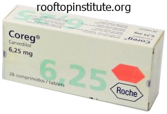
Order 12.5 mg coreg free shipping
The pleural thickening is usually most marked at the lung bases blood pressure chart spreadsheet coreg 6.25 mg purchase on-line, with obliteration of the costophrenic angles pulse pressure 31 trusted coreg 6.25 mg. When the pleural thickening is in depth, it causes restrictive ventilatory defect, which may give rise to dyspnoea. Epidemiology research show an elevated risk of lung cancer in employees in the asbestos industry, with an approximately linear relationship between the dose of asbestos and the occurrence of lung most cancers. The medical features, distribution of cell types, investigation and treatment of asbestos-related lung cancers are the identical as for those not related to asbestos exposure (see Chapter 12), but impairment of lung function because of asbestosis could preclude surgical procedure. There is pleural thickening in both mid zones, with some blunting of the costophrenic angles. Video-assisted thoracoscopic pleural biopsy may be needed for definitive histopathological diagnosis, and pleurodesis may be carried out at the similar time for management of a pleural effusion. As the tumour progresses, it encases the lung and may involve the pericardium and peritoneum, and can give rise to bloodborne metastases. Radical surgical procedure within the type of extrapleural pneumonectomy has been attempted but has generally not proved profitable. Radiotherapy is usually employed in an try and reduce the chance of spread of the tumour through biopsy tracks and may relieve pain. Chemotherapy, using drugs such as pemetrexed and cisplatin, leads to tumour shrinkage in about 40% of patients, though its influence on survival is unclear. Unfortunately, prognosis is poor, with most sufferers dying inside 2 years of prognosis. The death of a patient with a suspected occupational lung illness ought to be reported to the related authority, such as the coroner, who might wish to undertake a post mortem examination. Occupational lung illness 191 � Occupational asthma accounts for 10�15% of all instances of bronchial asthma in adults. Evidence based guidelines for the prevention, identification, and management of occupational asthma. The most likely diagnosis is: A mesothelioma B benign asbestos pleurisy C lung carcinoma D diffuse pleural thickening E pleural plaques 14. He has smoked 5 cigarettes/day for 20 years, has a pet cat and works in the aerospace trade. The more than likely diagnosis is: A berylliosis B tuberculosis C sarcoidosis D lung most cancers E occupational bronchial asthma A 68-year-old man presents with a 2-month history of increasing breathlessness and left-sided chest ache. He has just retired from his own profitable electrical business � a enterprise he set up on the age of 22 after qualifying as an electrician within the shipyards. The most probably analysis is: A pneumonia B benign pleural thickening C asbestosis D mesothelioma E lung most cancers 14. The patient has had heavy exposure to asbestos, which was used for pipe lagging in shipyards. Asbestos causes calcified pleural plaques (but not calcification of mediastinal nodes). There is a considerably increased risk of tuberculosis in sufferers with silicosis. Once the worker has developed sensitisation to an agent, additional publicity might provoke an early asthmatic response (reaching a peak within 30 minutes), a late asthmatic response (occurring 4�12 hours later) or a dual response. Once asthma turns into established, symptoms could persist even when away from the work surroundings. The X-ray suggests either benign asbestos pleural illness (thickening/effusion) or mesothelioma. The thrombus typically develops in the deep veins of the legs and then travels to the lungs, inflicting obstruction of the pulmonary vasculature. Pulmonary embolism is particularly common when thrombosis occurs in the proximal femoral or iliac veins and is much less more likely to occur when thrombosis is confined to the calf veins. Most pulmonary emboli come up within the deep veins of the legs, but they may often arise from thrombus within the inferior vena cava or the right facet of the heart, or from indwelling catheters in the subclavian or jugular veins. However, thrombosis in the deep veins of the leg, pelvis or abdomen could additionally be utterly silent. Deep vein thrombosis Factors predisposing to venous thrombosis had been described by Virchow as a triad of venous stasis, damage to the wall of the vein and hypercoagulable states: � Venous stasis occurs on account of immobility. Clinical features the scientific options of pulmonary embolism depend upon the size and severity of the embolism, as Respiratory Medicine Lecture Notes, Ninth Edition. In acute huge pulmonary embolism, the picture is usually of a patient recovering from latest surgery who suddenly collapses. Occlusion of a giant a half of the pulmonary circulation produces a catastrophic drop in cardiac output and the affected person collapses with hypotension, cyanosis, tachypnoea and engorged neck veins. Sometimes, the presentation is extra subacute, as a collection of Massive pulmonary embolism Acute >50% occlusion of circulation Sudden circulatory collapse. D-dimer is a breakdown product of crosslinked fibrin and levels are elevated in patients with thromboembolism. However, levels are also typically elevated in different hospitalised sufferers, so that D-dimer assays can be used to exclude, however to not confirm, venous thromboembolism. For example, a young girl on oral contraception who presents with isolated pleuritic pain is very unlikely to have pulmonary embolism if her respiratory price is beneath 20/min and chest X-ray, arterial blood gases and D-dimer are normal. She can be reassured without the need for admission to hospital or additional investigation. Prompt recognition and therapy of an acute minor embolism may forestall the occurrence of an enormous embolism. Chronic thromboembolic pulmonary hypertension is a situation by which recurrent emboli progressively occlude the pulmonary circulation, giving rise to progressive dyspnoea, pulmonary hypertension and right heart failure. Pulmonary embolism is each under- and overdiagnosed in clinical apply, resulting in some sufferers failing to obtain therapy for a potentially life-threatening condition and others being unnecessarily subjected to the risks of anticoagulant remedy. Dyspnoea, tachypnoea (respiratory fee >20/min) and pleuritic ache are the three cardinal options of pulmonary embolism. If none of these options is current, a prognosis of pulmonary embolism could be very unlikely. Investigations General investigations General investigations may yield clues that point towards a diagnosis of pulmonary embolism. They are significantly helpful in excluding different diagnoses: � Chest X-ray is usually regular, however elevation of a hemidiaphragm and areas of linear atelectasis may be seen on account of pulmonary emboli. A small pleural effusion with wedge-shaped peripheral opacities may happen in affiliation with pulmonary infarction, and an space of lung infarction may not often bear cavitation. In large embolism, an space of underperfusion with few vascular markings may be obvious. Enlarged pulmonary arteries are a feature of pulmonary hypertension in continual thromboembolic disease. The chest X-ray helps exclude different diagnoses, corresponding to pneumothorax, pneumonia and pulmonary oedema. The angiographic options of embolism are intraluminal filling defects, abrupt cut-off of vessels, peripheral pruning of vessels and areas of reduced perfusion. Very fast spiral images are obtained in the course of the injection of iodinated distinction medium right into a peripheral vein. It has higher specificity than ventilation�perfusion isotope scanning in the prognosis of pulmonary embolism, although it does involve a higher radiation dose.
Syndromes
- Burns
- VCUG (voiding cystourethrogram), a special x-ray to make sure the bladder is working
- Dairy products or food containing mayonnaise (such as coleslaw or potato salad) that have been out of the refrigerator too long
- Sunken eyes
- Creating an environment with low sensory stimulation
- Bleeding from a blood vessel or aneurysm in the brain (subarachnoid hemorrhage)
- Resting as much as possible for at least a week
Coreg 25 mg purchase line
Keywords: Arteriovenous shunts hypertension 7th buy coreg 6.25 mg on-line, Glomus body blood pressure chart for infants coreg 25 mg buy generic line, temperature regulation 25 the answer is D: Postcapillary venules within the subpapillary plexus. These histopathologic findings recommend a prognosis of cutaneous necrotizing vasculitis. Damage to the vessel wall is related to fibrin deposition and extravasation of erythrocytes (shown within the image). The lesions often develop in the postcapillary venules that drain from capillary loops to the subpapillary venous plexus. Postcapillary venules are identified to regulate the motion of inflammatory cells from the vascular house to regions of tissue damage during irritation (see Chapters 5 and 9). Cutaneous necrotizing vasculitis may be primary (without a recognized cause) or secondary 108 Chapter 8. None of the other blood vessels are principal targets for immune complex�mediated irritation in sufferers with cutaneous necrotizing vasculitis. Scleroderma is a chronic autoimmune disease characterised by excessive collagen deposition in the pores and skin and internal organs. Large, tightly packed collagen bundles parallel to the pores and skin surface are a characteristic histologic discovering in sufferers with scleroderma. Sweat glands and hair follicles are eventually destroyed, owing to the dearth of enough arterial blood supply. The hypodermis (choice B) is normally not concerned in early levels of this disease. Keywords: Scleroderma, dermis, reticular dermis 27 the answer is D: Hair follicles. Hair is present over most of the physique surface, apart from the pores and skin of the palms, soles, lips, glans penis, clitoris, and labia minora. Hair follicles are invaginations of the epidermis that reach from the surface of the dermis to the deep reticular dermis and/or hypodermis. In this low-magnification image, transverse sections of several hair follicles exhibit epidermal epithelial cell sheaths surrounding centrally situated hair shafts. Sebaceous glands (choice E) are related to hair follicles (arrowheads, shown in the image). The dermal papilla is continuous with the surrounding dermal connective tissue and incorporates a capillary community that nourishes and sustains the dwelling hair follicle. Hair matrix (choice E) refers to the layer of epithelial cells covering the dermal papilla. External root sheath (choice B) is a continuation of the dermis that covers the hair follicle (indicated by arrows in the image). Down progress of epidermal epithelial cells (particularly the basal and spinous layers) into the dermis is referred to as the exterior root sheath. The invaginating dermis is accompanied by its basement membrane deep into the dermis. Here, the basement membrane turns into thickened and is referred to as the "glassy membrane. The glassy membrane separates the hair follicle from the surrounding connective tissue sheath. None of the other cutaneous structures are steady with the glassy membrane of the hair follicle. Epithelial cells surrounding the dermal papilla are collectively referred as the hair matrix. Matrix cells characterize the proliferative (germinative) layer of the hair follicle, comparable to cells of the stratum basale of the dermis. Continuous proliferation and division of those cells account for the growth of hair. Melanocytes are scattered among the many matrix cells and move melanin to the developing hair cells, thereby offering the hair with its pure colour. Matrix cells also differentiate to form cells making up the interior root sheath (choice D). The inside root sheath consists of multiple cell layers and completely covers the preliminary a half of the hair shaft. Dermal papilla (choice A) consists of connective tissue whose primary function is to provide the vascular supply to the rising hair follicle. Inductive interactions between dermal papillae and matrix cells within the hair bulb are believed to regulate the dimensions and form of hair. Arrector pili are small bundles of clean muscle fibers extending from the midpoint of the external root sheath to the papillary dermis. Contraction of these muscle tissue causes hair shafts to become erect resulting in the formation of tiny skin bumps (so-called goose bumps). None of the opposite cutaneous constructions exhibit the distinctive histologic options of arrector pili muscle. The region of the external root sheath close to the insertion site of the arrector pili muscle and the origin of the sebaceous gland is referred to because the follicular bulge. This small structure is house to clusters of undifferentiated epithelial stem cells. Here, they proliferate and differentiate to resurface (regenerate) the skin lesion. Keywords: Epidermal stem cells, hair follicles, follicular bulge 33 the reply is D: Sebaceous glands. Sebaceous gland secretions enhance greatly at the time of puberty beneath the influence of circulating sex hormones. These hormonal changes also induce abnormal cornification of epithelial cells close to the neck of the sebaceous glands. Together, (1) excessive manufacturing of sebum (secretory product of sebaceous glands) and (2) irregular cornification result in the dilation of the sebaceous follicle. This bacterium proliferates in the affected glands, leading to an intense persistent inflammatory response. A quick duct delivers the secretory merchandise of a quantity of acini to the higher portion of the hair follicle. Sebaceous glands develop as an outgrowth of the external root sheath of the hair follicle (shown in the image). Undifferentiated, flattened epithelial cells encompass the periphery of glandular acini. The epithelial cells continually proliferate, move towards the middle of the acinus, and differentiate into massive lipid-producing cells with fat droplets of their cytoplasm. Sebum, the oily product of the sebaceous gland, is released following rupture of the cells. Sebum has antimicrobial and proinflammatory actions and serves to shield the stratum corneum of the epidermis. Keywords: Sebaceous glands, sebum 35 the reply is E: Secretory portion of eccrine sweat glands. Eccrine sweat glands are epidermal appendages that are independent of hair follicles.
Coreg 6.25 mg buy generic online
Rather arrhythmia names coreg 6.25 mg buy cheap on line, they characterize the fusion of precursor cells derived from mononuclear progenitor cells within the bone marrow blood pressure normal value coreg 12.5 mg cheap without prescription. Osteopetrosis, also referred to as "marble bone" illness or Albers-Sch�nberg disease, is a bunch of rare inherited problems. The sclerotic skeleton in sufferers with osteopetrosis is the end result of failed osteoclast operate. Because osteoclast function is arrested, osteopetrosis is characterised by block like radiodense bones, hence the term marble bone illness. Keywords: Osteopetrosis, Albers-Sch�nberg illness, osteoclasts 38 the answer is D: Increased osteoclast activity. Osteoporosis is the commonest bone illness characterised by progressive lack of bone mass. Porous bone tissue can now not provide enough mechanical support and eventually leads to bone fracture. Bone loss is the result of aggressive bone resorption by increased osteoclast exercise and decreased bone deposition by osteoblasts (therefore, not choices A, C, and E). Keywords: Osteoporosis, osteopenia fifty three 39 the reply is C: Impaired mineralization of new bone matrix. Osteomalacia (soft bones) is a disorder of adults characterized by inadequate mineralization of newly fashioned bone matrix (thus selection E is incorrect). Broad (exaggerated) layers of osteoid rim the bony trabeculae in patients with osteomalacia. Decreased bone formation by osteoblasts (choice A) and enhanced bone resorption by activated osteoclasts (choice B) are usually observed in sufferers with osteoporosis. Abnormal metabolism of vitamin D and/or inadequate calcium in the diet is the first cause of osteomalacia in adults and rickets in kids. Malabsorption of vitamin D and calcium complicates a selection of small intestinal diseases including celiac sprue, Crohn disease, and scleroderma. Insufficient amount of vitamin C (choice C) causes scurvy, in which collagen assembly is disrupted. The higher and lower limb bones of the appendicular skeleton develop by way of the process of endochondral ossification. Neural crest� derived flat bones of the cranium, face, and clavicle are fashioned by way of a different course of referred to as intramembranous ossification. Intramembranous ossification is a mechanism whereby bone is immediately developed on the embryonic mesenchymal tissue, the membrane. Osteoblasts derived from mesenchymal cells deposit bone matrix at a number of places and form a number of bone spicules. These bone spicules undergo remodeling and ultimately form compact bone at both the external surfaces and stay as spongy bone throughout the bone cavity. Keywords: Intramembranous bone formation 42 the reply is B: Chondrocytes within the proliferation zone of the growth plate. Chondrocytes within the proliferation zone of the expansion plate endure mitosis and type longitudinal columns of stacked cells, a specialized form of isogenous groups. The stacked cells produce and deposit new cartilage matrix to enhance the length of long bones throughout development. Chondrocytes within the hypertrophy zone (choice A) undergo apoptosis and no longer produce matrix. Chondrocytes within the resting zone (choice C) present reserve cells for proliferation. Keywords: Endochondral bone formation, development plate 54 Chapter 4 forty three the reply is B: Arrest of the epiphyseal development plate. Achondroplasia refers to a syndrome of short-limbed dwarfism and macrocephaly and represents a failure of normal epiphyseal cartilage formation. It is the commonest genetic type of dwarfism and is inherited as an autosomal dominant trait. This mutation negatively regulates chondrocyte proliferation and differentiation and arrests the event of the growth plate. The other steps of endochondral bone formation (choices A, C, D, and E) appear to be regular in patients with achondroplasia. Keywords: Achondroplasia, endochondral bone formation, growth plate forty four the reply is E: Lack of defect in intramembranous bone formation. Epiphyseal growth plates in patients with achondroplasia exhibit attenuated proliferation zones and reduced zones of proliferating cartilage. Thus, endochondral bone formation is impaired, while intramembranous bone formation proceeds normally. For this cause, bones that develop by way of the intramembranous ossification pathway, corresponding to flat bones of the neurocranium, seem relatively massive. As a result, patients with achondroplasia appear to have brief and thick limb bones. Keywords: Achondroplasia, intramembranous ossification forty five the answer is C: Area three. Epiphyseal plates are cartilaginous discs that lay between the diaphysis and the epiphysis of lengthy bones. From epiphysis to diaphysis, they include zones of resting cartilage (choice A), proliferation (choice B), hypertrophy (choice C), calcification (choice D), and ossification (choice E). Chondrocytes in the zone of hypertrophy (area 3) are tremendously enlarged, and the matrix positioned between columns of hypertrophic chondrocytes is compressed into thin septae. Keywords: Epiphyseal progress plate, endochondral bone formation 46 the reply is B: Area 2. Chondrocytes within the zone of proliferation endure a quantity of rounds of mitotic division. Cells throughout the same isogenous group are organized into longitudinal columns, and so they actively deposit a new cartilage matrix. Owing to the longitudinal proliferation of chondrocytes in the proliferation zone, the structure of the expansion plate stays essentially unchanged from early fetal life to skeletal maturity. The price of mitosis and manufacturing of latest cartilage matrix matches the velocity of ossification from the diaphysis. Keywords: Epiphyseal growth plate, endochondral bone formation 47 the reply is A: Cartilage progress plates. The growth of the width of the vertebrae is from appositional development of periosteum, a form of intramembranous ossification. Columns of chondrocytes in longitudinal isogenous teams actively deposit new bone matrix and lead to longitudinal development of vertebrae. Asymmetric growth of the cartilage development plates seems to be brought on primarily by an underlying asymmetry in the rate of cell cycle development in affected chondrocytes. Keywords: Scoliosis, progress plates forty nine the reply is A: Cartilage and dense connective tissue. During the therapeutic means of a bone fracture, necrotic particles and extravasated blood are removed by neutrophils and macrophages. Blood vessels and fibroblasts then proliferate at the website of damage to type a granulation tissue embedded in unfastened connective tissue. Keywords: Bone fracture, callus the fibrocartilaginous, soft 50 the answer is E: Woven.

Generic 12.5 mg coreg with visa
The corona radiata stays hooked up to the oocyte after ovulation and may not disperse until fertilization arteria appendicularis buy coreg 6.25 mg on line. The eosinophilic debris inside the antrum of this secondary follicle represents proteins within the liquor folliculi that precipitated during tissue fixation arrhythmia in 7 year old cheap coreg 12.5 mg amex. In this section, the zona pellucida appears as a skinny, eosinophilic ring between the oocyte and the corona radiata. These glycoproteins form a clear fibrous matrix that envelops the oocyte and early cleavage-stage embryo. The zona pellucida is degraded prior to implantation by proteolytic enzymes that are secreted by the blastocyst. The zona pellucida that surrounds the oocyte offers a multivalent array of receptors for sperm adhesion and activation of the sperm acrosome response. Theca externa cells (zone 4) embody fibroblasts and clean muscle cells (fibromuscular connective tissue). Primary oocytes complete the primary meiotic cell division a few hours before ovulation. One set of chromosomes is retained by the secondary oocyte, whereas the other set is discarded, together with a small amount of cytoplasm, as the primary polar physique. This large vesicle forms in the perivitelline house, between the germ cell plasma membrane and the zona pellucida. Granulosa cells synthesize aromatase, an intracellular enzyme that converts androstenedione into estradiol. Granulosa lutein and theca lutein cells (choices B and E) synthesize estrogen and progesterone during the secretory part of the menstrual cycle, after formation of the corpus luteum. Keywords: Ovaries, granulosa cells Female Reproductive System and Breast 17 the answer is D: Secondary oocyte. The resulting secondary oocyte is characterized by the presence of a single (first) polar physique. During ovulation, the secondary oocyte is launched from the floor of the ovary and enters the ampulla of the uterine tube. Zygote (choice E) describes the postfertilization embryo, prior to the primary cleavage division. Keywords: Ovaries, secondary oocytes, ovulation 18 the reply is A: Fertilization. The biochemical mechanisms that regulate this complex process are poorly understood. None of the other cellular or physiological processes regulate the second meiotic division. Keywords: Fertilization, meiosis 19 the answer is B: Formation of the second polar body. Fertilization is a posh developmental process that brings collectively haploid gametes, restores a diploid genome, and sets in motion early improvement. Binding of sperm to the zona pellucida triggers the sperm acrosome response (choice D). The acrosome response liberates hydrolytic enzymes that disperse the corona radiata (choice A) and create openings in the zona pellucida that facilitate the entry of hypermotile sperm into the perivitelline space. Movements of male and female pronuclei (choice C) could be difficult to monitor by gentle microscopy. On the other hand, the formation of a second polar body can be monitored easily utilizing an inverted section microscope. Formation of the second polar physique provides unambiguous evidence of fertilization. It signifies completion of the second meiotic division and heralds the initiation of early development. The postfusion oocyte undergoes a speedy sequence of changes that block the entry of more than one spermatozoan. Three postfusion blocks to polyspermy have been described: (1) membrane depolarization (fast block), (2) Ca2+��mediated cortical granule release (cortical reaction), and (3) enzymemediated modification of the zona pellucida (zona reaction). Exocytosis of cortical granules liberates proteases that degrade sperm receptors on the oocyte plasma membrane and on the zona pellucida. Mitochondrial membrane permeability transition (choice E) regulates programmed cell demise. Atretic follicles endure degenerative changes related to programmed cell dying (apoptosis). Macrophages enter atretic follicles to remove mobile debris, and fibroblasts produce collagen scar tissue that may resorb over time. The circle proven on this image reveals a small cystic cavity surrounded by pink collagen scar tissue. This part also reveals stratification of the theca folliculi that surrounds a secondary (antral) follicle. This picture exhibits the stays of an atretic follicle that has been invaded by stromal connective tissue. Most of the mobile debris have been removed by macrophages; nevertheless, the glycoproteins that comprise the zona pellucida are resistant to degradation. The remnant of a zona pellucida is visible on this image as an eosinophilic loop that resembles a folded rubber band. This fibrous construction is situated in a cystic area that was occupied by the primary oocyte. Recent studies indicate that follicular atresia is triggered by down-regulation of an apoptosis-inhibitory protein in granulosa cells. Without this inhibitory protein, granulosa cells exit the cell cycle and activate hydrolytic enzymes. Death of the oocyte ensues, and the follicle is infiltrated by macrophages that take away apoptotic bodies and necrotic tissue debris. Condensation of chromatin (pyknosis), degradation of chromatin (karyolysis), and eosinophilic apoptotic our bodies present evidence of irreversible cell damage. Examination of the ovaries in the pathology department would therefore reveal proof of necrosis and apoptosis, as well as evidence of healing (collagen scar 248 Chapter 17 tissue). Atrophy of the ovaries happens in postmenopausal woman, owing to the lack of hormonal stimulation. Germ cell tumors (choice A), endometriosis (choice C), ovarian cancer (choice D), and secondary lymphoid nodules (choice E) could affect the ovaries; however, these pathologic situations are less frequent than normal patterns of scar tissue. After ovulation, the stratum granulosum collapses and varieties a temporary endocrine organ that secretes progesterone and estrogen. This steroid hormone factory is referred to because the corpus luteum (L: yellow body).

25 mg coreg order fast delivery
Pillar cells contain bundles of keratin that make the cells stiff to define the tunnel of Corti arteria meningea anterior purchase coreg 25 mg line. Sulcus spiralis internus (choice D) represents the concavity created by the inside projection of the spiral limbus (right side of the image) arrhythmia episode coreg 6.25 mg purchase with visa. The tectorial membrane hangs over this space to attain the spiral organ, thereby creating a tunnel-like space 285 (referred to as the interior spiral tunnel). The spiral organ of Corti is a highly specialised epithelium resting on the basilar membrane and uncovered to the endolymph within the scala media. Hair cells are particular auditory receptors and sensory transducers that detect the amplitude and frequency of sound waves. There are two types of hair cells in the spiral organ, particularly inner and outer hair cells. The inner hair cells (choice B) kind a single row of cells alongside the inside pillar cells. The outer hair cells are organized into three rows at the base of the cochlea (as shown in this specimen) and enhance to five rows at the apex. Phalangeal cells (choice D) and pillar cells (choices E, indicated by arrowheads) present help to the hair cells. The outer phalangeal cells can be distinguished from the outer hair cells by their location in this image (the three well-aligned nuclei instantly under the three outer hair cells). Hensen cells (choice A) are exterior limiting cells on the lateral facet of the spiral organ. Keywords: Ears, spiral organ of Corti, hair cells fifty two the answer is B: Oval window. The oval window and round window are two openings of the bony labyrinths inside the temporal bone. The oval window is situated on the lateral wall of the vestibule of the bony labyrinth. Movement of the stapes induced by the vibration of the tympanic membrane stirs up the mechanical vibration of the perilymph contained in the scala vestibuli, which in flip causes vibration of the endolymph within the scala media and, subsequently, the perilymph within the scala tympani. The round window (choice C) is located at the inferior facet of the bottom of the cochlea and is roofed by an elastic membrane termed secondary tympanic membrane. Pressure modifications of fluid in the cochlea trigger movement (bulging out or in) of this membrane. None of the opposite structures mediate sound wave conduction from the middle ear to the internal ear. Keywords: Sound conduction, ears, oval window fifty three the reply is A: Basilar membrane. As sound vibrations are transferred to the inner ear, a stress pulse of the perilymph of the scala vestibule causes a touring wave of deformation alongside the basilar membrane. The traveling wave of sound of a particular frequency reaches its peak amplitude at a specific location alongside the basilar membrane. As mentioned earlier, the basilar membrane is 286 Chapter 19 slim and comparatively stiff on the base of the cochlea but increases in width and reduces in stiffness because it coils toward the apex of the cochlea. High-frequency sounds cause maximal amplitude of the basilar membrane near the base of the cochlea. By distinction, the basilar membrane close to the apex of the cochlea undergoes maximal displacement in response to low-frequency sounds. Thus, totally different sites alongside the basilar membrane are particular for sounds with particular frequencies (pitch) and provide a structural foundation for frequency discrimination. The receptor cells of the organ of Corti resting on a particular web site of the basilar membrane respond finest to sounds at explicit frequency and convert the mechanical tuning of the basilar membrane into nerve pulses. The diploma of displacement of the basilar membrane, in one other phrases, the amplitude at any specific frequency, displays the depth or loudness of sound. None of the other structures encode acoustic information based mostly on sound frequency or amplitude. Keywords: Ears, basilar membrane 54 the answer is C: Hair cells of the spiral organ of Corti. The receptor hair cells of the organ of Corti are supported and surrounded by phalangeal cells. At their apical floor, stereocilia of the hair cells attach to the tectorial membrane. The basilar membrane stretches from the osseous spiral lamina medially to the lateral spiral ligament, whereas the tectorial membrane hinges from the spiral limbus. Vibrations of the basilar membrane and tectorial membrane create a shearing impact that deflects and prompts stereocilia of the hair cells. The activated hair cells generate action potentials that are conveyed by the cochlear nerve to the central nervous system. Hair cells of the crista ampullaris and macula (choices A and B) are receptor cells liable for balance and equilibrium. Keywords: Sound perception 55 the reply is B: Dilation of the endolymphatic system. M�ni�re illness is the triad of vertigo, sensorineural hearing loss, and tinnitus. M�ni�re illness is characterized pathologically by hydropic distention of the endolymphatic channels of the membranous labyrinth. Dilation of the cochlear duct and saccule occurs at the early stage of illness, and ultimately, the whole endolymph-containing network of channels is involved. Patients are afflicted with in depth vertigo and tinnitus, accompanied by nausea and vomiting. None of the other mechanisms of illness are related to the pathogenesis of M�ni�re disease. Various organs and tissues are examined in the course of the autopsy of a 70-year-old girl. The wound is cleaned and sutured; however, the boy suffers temporary lack of sensation distal to the wound. Which of the photographs proven above represents an example of a tissue that may be expected to present degenerative changes in the injured finger of this patient The sections proven below represent 4 completely different parts of the nervous system. The five sections shown beneath had been obtained from cell-rich glandular tissues which are organized into clusters, acini, or cords. Various lymphoid organs are examined at low magnification within the histology laboratory. Various portions of the digestive tract are examined by gentle microscopy at low magnification. Various endocrine and reproductive organs are examined at low magnification within the histology laboratory. You study the biopsy and observe a quantity of normal structures in a region adjacent to the neoplasm (shown in the image). A transverse part through the posterior facet of this organ is examined by mild microscopy (shown in the image).
Chromium Proteinate (Chromium). Coreg.
- You have a chromate allergy.
- What is Chromium?
- Type 2 diabetes.
- Dosing considerations for Chromium.
- Are there safety concerns?
Source: http://www.rxlist.com/script/main/art.asp?articlekey=96895
Generic coreg 12.5 mg on-line
Myelin surrounding nerve fibers within the white matter of the brain and spinal twine is composed of concentric layers of oligodendrocyte cell membrane blood pressure over 60 6.25 mg coreg purchase mastercard. Multiple heart attack one direction buy 25 mg coreg with visa, tongue-like processes of particular person oligodendrocytes are capable of myelinate a quantity of nearby axons. Schwann cells (choice E) produce myelin for axons within the peripheral nervous system. In this teased preparation of a peripheral nerve, the axon is visible as the darkish red line throughout the myelinated nerve fiber. Axons (also referred to as nerve fibers) are cell processes that conduct action potentials away from neuronal cell our bodies. In this slide preparation, the myelin sheath was eliminated by solvent extraction and seems as empty (or foamy) house surrounding the axons. Numerous Schwann cell nuclei (choice E) are observed on this image residing along the myelin sheath. Long axons inside a peripheral nerve are myelinated by multiple Schwann cells, leading to a segmented myelin sheath. The junction where two successive Schwann cells meet is referred to as a node of Ranvier, and the myelin sheath between two sequential nodes of Ranvier is referred as an internodal segment (choice B). Thus, a 1-m nerve leaving the spinal cord is associated with over 1,000 Schwann cells. Schmidt-Lanterman clefts (choice E), also termed myelin clefts, are areas containing Schwann cell cytoplasm that lies between adjoining myelin membrane lamellae. Schmidt-Lanterman clefts, endoneurium, and perineurium (choices E, A, and D) are additionally not seen on this preparation. Neuronal cell bodies that cluster together within the peripheral nervous system are referred to as ganglia. Satellite cells in dorsal root ganglia form an entire monolayer surrounding the central neurons. Keywords: Dorsal root ganglion, satellite tv for pc cells 23 the answer is E: Pseudounipolar. They possess a single axon that leaves the neuronal cell physique and varieties two axonal branches: peripheral and central. These neurons are described as being pseudounipolar Nerve Tissue as a outcome of their two axonal processes arise from a single process. Interneurons and motor neurons (choice C) are both multipolar neurons that include one axon and a varied variety of dendrites (dendritic tree). Keywords: Dorsal root ganglion, pseudounipolar neuron 24 the answer is E: Visceral sensory. Visceral motor and autonomic motor neurons (choices A and D) usually provide motor innervation to the viscera and glands; their postsynaptic neurons are located in the sympathetic or parasympathetic ganglia. Multipolar neurons located within the ventral horn of the spinal twine provide somatic motor innervation to skeletal muscle (choice C). Astrocytes are identified as giant cells with "stellate" processes projecting in all directions. Proliferation of astrocytes and microglia in response to damage is referred to as gliosis. Gliosis within the central nervous system is the equal of scar formation elsewhere within the physique. However, after 2 weeks, gliosis has taken place and makes an attempt at axonal regeneration come to an finish. Axonal regeneration seems to require intimate contact with extracellular fluids containing plasma proteins. Keywords: Cerebral contusion, gliosis, astrocytes 26 the answer is E: Support and nourishment of neurons. Fibrous astrocytes are primarily positioned in the white matter and have relatively few however long ninety one processes. Some of the processes could be very long and even span the whole thickness of the mind. The expanded "perivascular toes" of some of the processes of protoplasmic astrocytes terminate on capillaries and contribute to the formation of the "blood�brain barrier. Defense against pathogens and phagocytosis (choices A and D) are essential features of microglial cells. Myelination of axons (choice C) and production of cerebrospinal fluid (choice B) are the principal functions of oligodendrocytes and ependymal cells, respectively. In areas of harm or infectious illness, microglia accumulate in massive numbers and demonstrate an infiltrative phenotype with attribute thin elongated nuclei. This distinctive nuclear morphology makes them readily seen in routine H&E preparations (shown in the image). They are derived from monocytes that migrate into the brain as it turns into vascularized during embryonic and fetal development. Glioblasts (choice A) and neuroblasts (choice D) are precursors for macroglial cells and neurons, respectively. These stem cell populations are derived themselves from neuroepithelial cells (choice E) which might be present in the wall of the neural tube throughout early growth. Microglia play crucial function in protection in opposition to infectious microorganisms and neoplastic cells. As macrophage-like phagocytic cells, they function to remove injured cells and necrotic debris. Keywords: Microglia, phagocytosis ninety two Chapter 7 30 the reply is D: Oligodendrocytes. Concentric layers of oligodendrocyte plasma membrane type the myelin sheath surrounding axons. Thus, a single oligodendrocyte can kind a number of internodal segments surrounding a number of neuronal axons. On routine H&E-stained slides, oligodendrocytes exhibit a memorable "fried egg" appearance with central, small, dark spherical nuclei surrounded by a halo of vacuolated cytoplasm. The ciliated, cuboidalcolumnar cells that line the mind ventricles and spinal canal are termed ependymal cells. The epithelial-like ependymal cell layer types a barrier between the cerebrospinal fluid and mind parenchyma and regulates fluid transport between these two compartments. At particular areas throughout the ventricular system, ependymal cells are modified to kind a choroid plexus with related capillaries that secrete cerebrospinal fluid. None of the other cells listed as choices displays a low columnar epithelial morphology. Nerve axons, along with their myelin sheaths and associated Schwann cells, are enveloped inside several layers of connective tissue. The outermost irregular connective tissue layer wrapping a peripheral nerve is termed epineurium. Epineurium is misplaced near the termination of nerve fibers with free nerve endings however contributes to the capsules of encapsulated nerve endings. The collagen bundles within the epineurium are largely responsible for the remarkable tensile power of peripheral nerves. Nerve fascicles are surrounded by perineurium-a skinny sheath composed of concentric layers of specialized epithelial-like connective tissue cells.
Coreg 12.5 mg buy on-line
The circle shown in the picture identifies a portal triad composed of a portal vein heart attack quiz questions buy cheap coreg 25 mg line, bile duct blood pressure medication to treat acne coreg 12.5 mg order mastercard, and hepatic artery. The portal vein (choice E) is thin walled, and its diameter is way larger than that of the hepatic artery (choice C). It delivers poorly oxygenated, but nutrient-rich, blood to hepatocytes lining the sinusoids. Hepatic arteries arise from the celiac trunk-an unpaired branch of the belly aorta. None of the other choices exhibit histologic options of the hepatic portal triad. This image reveals the central veins (terminal hepatic venules) of two adjoining liver lobules (arrows, shown within the image). They coalesce to type sublobular veins (choice D) that drain to hepatic veins that vacant into the inferior vena cava. Keywords: Liver, terminal hepatic venules four the reply is E: Glucuronyltransferase. In order to be removed from the circulation, bilirubin must be transported into hepatocytes, conjugated with glucuronic acid (to make it water soluble), after which excreted into the bile for elimination. Approximately 70% of regular newborns exhibit a transient unconjugated hyperbilirubinemia. This "physiological jaundice" is more pronounced in untimely infants due to insufficient hepatic clearance of bilirubin associated to organ immaturity. High concentrations of unconjugated bilirubin in a neonate can cause irreversible brain harm (referred to as kernicterus). Patients with right-sided heart failure have pitting edema of the lower extremities and an enlarged and tender liver. A generalized enhance in venous pressure, typically from chronic right-sided coronary heart failure, results in a rise in the quantity of blood in many organs. The liver is especially weak to persistent passive congestion as a outcome of the hepatic veins empty into the vena cava instantly inferior to the center. In patients with continual passive congestion of the liver, the central veins of the hepatic lobule turn into dilated. Increased venous pressure leads to dilation of the sinusoids and strain atrophy of centrilobular hepatocytes. The different decisions are less commonly affected by continual passive congestion of the liver. Keywords: Liver sinusoids, congestive coronary heart failure 6 the reply is D: Portal vein. Approximately 75% of the blood flowing via the liver is derived from the hepatic portal vein. Hepatic sinusoids are lined by a discontinuous endothelium that facilitates entry of hepatocytes to the blood. Moreover, the basal lamina of the endothelium is absent over massive areas, and there are gaps between adjoining cells. Hepatic sinusoids are also lined by resident macrophages (referred to as Kupffer cells). None of the opposite cytologic features characterize endothelial cells lining hepatic sinusoids. Keywords: Liver sinusoids, fenestrated capillaries 8 the reply is D: Space of Disse. Hepatocytes are separated from vascular endothelial cells and Kupffer cells by a perisinusoidal space (of Disse). This microscopic area supplies a location for the change of fluid and biomolecules between hepatocytes and blood. Microvilli on the hepatocyte basal membrane fill the space of Disse and improve the floor area obtainable for transport (endocytosis and exocytosis). RokitanskyAschoff sinuses (choice C) are deep invaginations of the mucosa in the wall of the gallbladder. The space of Mall (choice E) is positioned between hepatocytes and connective tissue of the portal triads. Hemosiderin is a partially denatured form of ferritin that aggregates easily and is recognized microscopically as yellow-brown granules throughout the cytoplasm. Hereditary hemochromatosis is an abnormality of iron absorption within the small intestine. In this genetic illness, iron is saved mostly within the form of hemosiderin, primarily within the liver. The liver is the principal organ concerned in detoxification of foreign substances, together with industrial chemical compounds, pharmaceutical medication, and bacterial toxins. Small doses of acetaminophen (an analgesic) are absorbed from the abdomen and small gut and conjugated in the liver to form unhazardous derivatives. In instances of overdose, the traditional pathway of acetaminophen metabolism is saturated. Excess acetaminophen is then metabolized in the liver via the blended function oxidase (cytochrome P450) system, yielding oxidative metabolites that cause predictable hepatic necrosis. These metabolites initiate lipid peroxidation, which damages the plasma membrane and leads to hepatocyte cell dying. The poisonous dose of acetaminophen after a single acute ingestion is in the range of a hundred and fifty mg/kg in children and 7 g in adults. None of the opposite enzymes metabolizes acetaminophen to generate reactive oxygen species. Keywords: Liver, predictable necrosis 197 eleven the reply is A: Blood merchandise from the spleen. The scattered black objects characterize Kupffer cells which have picked up carbon particles from the circulation. Their mobile processes span the hepatic sinusoids, searching for necrotic particles and international materials to ingest. Bile provides a car for the elimination of ldl cholesterol and bilirubin, and bile salts facilitate the digestion and absorption of dietary fats. Hepatocytes excrete bile into small canals (canaliculi) that drain to bile ducts throughout the portal triads. These cuboidal to columnar epithelial cells repeatedly monitor the composition and move of bile. The patient described on this clinical vignette has an autoimmune disease (primary biliary cirrhosis) that results in continual destruction of intrahepatic bile ducts. As a result of this damaging inflammatory course of, the small bile ducts all but disappear. Keywords: Primary biliary cirrhosis, cholangiocytes thirteen the answer is C: Hepatic stellate cells. Vitamin A is essential for imaginative and prescient, wholesome skin, and proper functioning of the immune system. These mesenchymal cells are located between hepatocytes and endothelial cells within the perisinusoidal area of Disse. They store vitamin A as retinyl esters and secrete retinol bound to retinol-binding protein.
Cheap coreg 6.25 mg fast delivery
Together prehypertension pubmed coreg 6.25 mg buy lowest price, thick insoluble cell and lipid envelopes kind an effective epidermal water barrier heart attack 42 year old buy 12.5 mg coreg fast delivery. Keywords: Epidermis, lamellar bodies 15 the reply is E: Slower degradation of melanin. The most important think about determining skin colour is the content material of melanin-a product of tyrosine metabolism. Melanocytes are neural crest�derived dendritic cells which are scattered amongst basal epidermal cells. They produce melanin within melanosomes and distribute these melanin granules to neighboring keratinocytes. The ratio of melanocytes to keratinocytes within the basal layer of the dermis is referred to because the epidermal melanin unit. This ratio differs between totally different elements of the body however is basically the identical in all races. Skin colour differences between the races are associated primarily to the destiny of melanin. In people with darker pores and skin, the degradation of melanin within lysosomes proceeds at a slower price than in light-skinned individuals. As a result of slower degradation in dark-skinned individuals, melanosomes are more broadly distributed throughout the epidermis. As a result of quicker degradation in light-skinned people, melanosomes are sparse within the higher layers of the epidermis. Increased number of melanocytes and elevated synthesis of melanin (choices B and C) are biological responses to ultraviolet radiation. Keywords: Melanocytes, melanosomes sixteen the reply is B: Increased manufacturing of melanin. Melanin absorbs ultraviolet radiation and protects the pores and skin from the harmful results of daylight. Harmful effects of daylight publicity on the skin embody inhibition of metabolism, technology of reactive oxygen species, activation of programmed cell death, mutagenesis, and most cancers. Cancers linked to ultraviolet radiation including basal cell carcinoma, squamous cell carcinoma, and melanoma. As a response to ultraviolet radiation exposure, melanocytes increase in cell quantity (hyperplasia) and speed up their production of melanin. None of the opposite organic processes are associated to the event of a suntan. If not recognized and removed at an early stage of tumor development, melanomas may be deadly. Acral lentiginous melanoma is the most typical sort of melanoma within the dark-skinned population. This melanoma usually arises within the palm of the hand, sole of the foot, or in a subungual (nail bed) location. During the early radial progress section, the tumor seems as an irregular, brown-to-black patch. As the disease progresses, neoplastic cells invade the deep dermal layer of the skin (vertical progress phase). Keywords: Melanoma, acral lentiginous, melanocytes 18 the answer is C: Langerhans cells. Langerhans cells are mononuclear phagocytes derived from bone marrow precursor cells. Monocytes in the bloodstream migrate into the epidermis the place they differentiate into Langerhans dendritic cells. Once an antigen is internalized, processed, and offered on the cell surface, Langerhans cells depart the epidermis and migrate to a regional lymph node. Here, Langerhans cells current foreign antigens to B and T lymphocytes (immunocytes). In routine H&E preparations, the nuclei of Langerhans cells stain intensely with hematoxylin, and the cytoplasm is obvious. Keywords: Langerhans cell histiocytosis, Langerhans cell 20 the reply is D: Merkel cell. The skin is the most important organ in the body and, as such, offers an intensive surface space for the processing exterior stimuli. A wealthy provide of terminal nerve fibers and a wide selection of sensory receptors are discovered within the dermis and dermis. Some myelinated afferent (sensory) nerve fibers lose their myelin sheath when penetrating the epidermal basement membrane. Uncontrolled malignant proliferation of Merkel cells is referred to as a Merkel cell carcinoma. Keywords: Mechanoreceptor, Merkel disk, Merkel cell 21 the answer is E: Pacinian corpuscle. Pacinian corpuscles are encapsulated tactile mechanoreceptors embedded within the deep dermis and hypodermis. They are giant oval "onion-like" buildings, with an outer capsule and numerous skinny, concentric lamellae. A extremely branched unmyelinated axon ending is current within the center of the corpuscle. Pacinian corpuscles are specialized for sensation of deep stress and transient vibrations via displacement of capsular lamellae. Free nerve endings (choice A) are axon terminals that lack a Schwann cell or connective tissue covering. Nerve fibers (choice D) are seen throughout the dermis as a set of myelinated nerve axons. None of the opposite selections describe the distinctive features of a pacinian corpuscle. Meissner corpuscles are encapsulated receptors specialized for the detection of shape and texture during energetic touch. These elliptical buildings are located within the cores of dermal papillae, with their long axis perpendicular to the surface of the pores and skin. The "twisted skein of wool" appearance is due 107 to a spiral arrangement of Schwann cells. Several unmyelinated endings of myelinated axons course via the Schwann cell corpuscle. Capillary loops (choice A) are found in dermal papillae; however, these tubular constructions usually exhibit a lumen. None of the opposite structures exhibit the distinctive features of a Meissner corpuscle. The pores and skin is richly equipped with blood via two major vascular plexuses: deep and superficial. Cutaneous arteries and veins traveling within the hypodermis type a deep subcutaneous (hypodermic) plexus. Ascending arterioles arising from arteries within the hypodermis cross the reticular dermis. Near the border of the papillary dermis, neighboring ascending arterioles anastomose to type a superficial or subpapillary plexus.
Coreg 6.25 mg cheap without prescription
It is a vital mechanism by which the physique terminates the action of many medication blood pressure value chart 6.25 mg coreg purchase with mastercard. Most medication are comparatively lipid-soluble as given blood pressure medication good for kidneys effective coreg 25 mg, a characteristic wanted for absorption throughout membranes. The similar property would lead to very slow removing from the body as a result of the unchanged molecule would also be readily reabsorbed from the urine within the renal tubule. The body hastens excretion by transforming many medication to much less lipid-soluble, much less readily reabsorbed forms. The transporter expels drug molecules from the cytoplasm into the extracellular house. Phase I Reactions Phase I reactions embody oxidation (especially by the cytochrome P450 group of enzymes, also known as mixed-function oxidases), discount, deamination, and hydrolysis. These enzymes are found in high concentrations within the easy endoplasmic reticulum of the liver. Nevertheless, some selectivity may be detected, and optical enantiomers, specifically, are often metabolized at completely different charges. The subgroups which are added embrace glucuronate, acetate, glutathione, glycine, sulfate, and methyl groups. Most of those teams are comparatively polar and make the product much less lipid-soluble than the unique drug molecule. For a number of medication, age or disease-related differences in drug metabolism are significant. Smoking is a typical cause of enzyme induction in the liver and lung and should enhance the metabolism of some medicine. Because the rate of biotransformation is usually the primary determinant of clearance, variations in drug metabolism should be thought of carefully when designing or modifying a dosage routine. Genetic Factors Several drug-metabolizing systems have long been recognized to differ among households or populations in genetically decided methods. Effects of Other Drugs Coadministration of sure agents may alter the disposition of many drugs. Enzyme induction-Induction (increased price and extent of metabolism) normally outcomes from elevated synthesis of cytochrome P450 drug-oxidizing enzymes within the liver in addition to the cofactor, heme. Several cytoplasmic drug receptors have been identified that result in activation of the genes for P450 isoforms. Many isozymes of the P450 household exist, and most inducers selectively enhance one or more subgroups of isozymes. Common inducers of some of these isozymes and the drugs whose metabolism is increased are listed in Table 4�3. Several days are normally required to attain maximum induction; an analogous period of time is required to regress after withdrawal of the inducer. The commonest robust inducers of drug metabolism are carbamazepine, phenobarbital, phenytoin, and rifampin. Enzyme inhibition-A few common inhibitors and the medication whose metabolism is diminished are listed in Table 4�4. Suicide inhibitors are medicine which are metabolized to merchandise that irreversibly inhibit the metabolizing enzyme. Such agents include ethinyl estradiol, norethindrone, spironolactone, secobarbital, allopurinol, fluroxene, and propylthiouracil. Metabolism may be decreased by pharmacodynamic factors such as a reduction in blood flow to the metabolizing organ (eg, propranolol reduces hepatic blood flow). P-gp inhibitors include verapamil, mibefradil (a calcium channel blocker no longer on the market), and furanocoumarin parts of grapefruit juice. Important medication which are normally expelled by P-gp (and are due to this fact probably extra poisonous when given with a P-gp inhibitor) include digoxin, cyclosporine, and saquinavir. This intermediate is conjugated with glutathione to a 3rd harmless product if glutathione stores are enough. If glutathione stores are exhausted, nonetheless, the reactive intermediate combines with sulfhydryl groups on important hepatic cell proteins, leading to cell dying. Prompt administration of different sulfhydryl donors (eg, acetylcysteine) could also be life-saving after an overdose. In severe liver disease, shops of glucuronide, sulfate, and glutathione may be depleted, making the affected person more susceptible to hepatic toxicity with near-normal doses of acetaminophen. You are planning to treat chronic major despair in a 35-year-old affected person with recurrent suicidal ideas. You are involved about drug interactions brought on by changes in drug metabolism on this patient. Reports of cardiac arrhythmias brought on by unusually excessive blood levels of two antihistamines, terfenadine and astemizole, led to their elimination from the market. Which of the following medicine might inhibit the hepatic microsomal P450 liable for warfarin metabolism Which of the following medicine, if used chronically, is most probably to enhance the toxicity of acetaminophen Which of the next drugs has larger first-pass metabolism in men than in women Which of the following drugs is a longtime inhibitor of P-glycoprotein (P-gp) drug transporters Which of the next cytochrome isoforms is liable for metabolizing the most important number of medicine This facilitates elimination of medicine that might in any other case be reabsorbed from the renal tubule. The clean endoplasmic reticulum, which incorporates the mixed-function oxidase drug-metabolizing enzymes, is selectively elevated by inducers. Rifampin and carbamazepine can induce drug-metabolizing enzymes and thereby might reduce the length of drug action. Displacement of drug from tissue may transiently improve the intensity of the impact however decreases the volume of distribution. Treatment with rifampin and continual alcohol use are related to increased drug metabolism and decrease, not greater, blood levels. Ketoconazole, itraconazole, erythromycin, and a few substances in grapefruit juice sluggish the metabolism of certain older non-sedating antihistamines (Chapter 16). Amiodarone is an important antiarrhythmic drug and has a well-documented capacity to inhibit the hepatic metabolism of many medication. Ethanol and sure different drugs induce P450 enzymes and thus cut back the hepatotoxic dose. Independent of physique weight and other factors, men have larger gastric ethanol metabolism and thus a lower ethanol bioavailability than women. Verapamil is an inhibitor of P-glycoprotein drug transporters and has been used to enhance the cytotoxic actions of methotrexate in most cancers chemotherapy. Know which P450 isoform is responsible for the greatest variety of necessary reactions. Describe the mechanism of hepatic enzyme induction and listing three medication that are known List 3 medication that inhibit the metabolism of other medicine. Describe some of the effects of smoking, liver illness, and kidney disease on drug elimination.

Buy generic coreg 12.5 mg
Choices involving chemical or physiologic antagonism are incorrect as a outcome of novamine is said to act on the identical receptors as acetylcholine blood pressure what is too low order 25 mg coreg fast delivery. When given alone blood pressure 200120 25 mg coreg cheap with amex, the novamine effect is opposite to that of acetylcholine, so choice C is inaccurate. Spare receptors affect the sensitivity of the system to an agonist because the statistical chance of a drug-receptor interplay increases with the total number of receptors. Similarly, no data on efficacy (maximal effect) is presented; this requires graded dose-response curves. Although each medicine are mentioned to be producing a therapeutic effect, no info on their receptor mechanisms is given. Which of the curves in the graph describes the percentage of binding of a large dose of full agonist to its receptors as the focus of a partial agonist is increased from low to very high ranges The binding of a full agonist decreases as the concentration of a partial agonist is increased to very high ranges. Curve 1 describes the response of the system when a full agonist is displaced by increasing concentrations of partial agonist. This is because the rising proportion of receptors binding the partial agonist lastly produce the maximal impact typical of the partial agonist. Partial agonists, like full agonists, bind one hundred pc of their receptors when present in a high sufficient focus. In contrast, pharmacologic antagonists bind to the agonist website and stop access of the agonist. The distinction may be detected experimentally by evaluating competition between the binding of radioisotopically labeled antagonist and the agonist. High concentrations of agonist displace or stop the binding of a pharmacologic antagonist but not an allosteric antagonist. Predict the effect of a partial agonist in a affected person within the presence and in the absence of Name the types of antagonists utilized in therapeutics. Describe the difference between an inverse agonist and a pharmacologic antagonist. Specify whether or not a pharmacologic antagonist is competitive or irreversible primarily based on its effects on the dose-response curve and the dose-binding curve of an agonist within the presence of the antagonist. Give examples of aggressive and irreversible pharmacologic antagonists and of Name 5 transmembrane signaling strategies by which drug-receptor interactions exert Describe 2 mechanisms of receptor regulation. A drug may have excessive efficacy however low efficiency or vice versa the flexibility to activate (agonism) or inhibit (antagonism) a biologic system or impact. In addition, the binding of a drug may be on the web site that an endogenous ligand binds that receptor, or at a different site Many medicine act on intracellular capabilities but reach their targets in the extracellular area. On reaching the goal tissue, some medicine diffuse via the cell membrane and act on intracellular receptors. Most act on receptors on the extracellular face of the cell membrane and modify the intracellular operate of these receptors by transmembrane signaling Receptors are in dynamic equilibrium, being synthesized within the interior of the cell, inserted into the cell membranes, sequestered out of the membranes, and degraded at varied rates. These adjustments are noted as upregulation or downregulation of the receptor numbers and usually take days to accomplish. The main processes involved in pharmacokinetics are absorption, distribution, and elimination. Appropriate utility of pharmacokinetic data and some easy formulas makes it attainable to calculate loading and maintenance doses. Units: liters the ratio of the rate of elimination of a drug to the focus of the drug within the plasma or blood. Units: volume/time, eg, mL/min or L/h the time required for the amount of drug in the physique or blood to fall by 50%. For drugs eradicated by first-order kinetics, this number is a continuing whatever the concentration. Units: time the fraction (or percentage) of the administered dose of drug that reaches the systemic circulation the graphic space under a plot of drug focus versus time after a single dose or throughout a single dosing interval. The plasma concentration is a function of the speed of input of the drug (by absorption) into the plasma, the rate of distribution, and the rate of elimination. These parameters are distinctive for a selected drug and a specific patient however have common values in giant populations that can be used to predict drug concentrations. Because the size of the compartments to which the drug may be distributed can vary with body size, Vd is sometimes expressed as Vd per kilogram of physique weight (Vd/kg). With 20 units of the drug within the body, the steady-state distribution leaves a blood concentration of 2 items. At equilibrium, only 2 items of the whole are current in the extravascular quantity, leaving 18 models nonetheless in the blood. In every case, the whole quantity of drug in the physique is similar (20 units), but the apparent volumes of distribution are very totally different. Drug C is avidly certain to molecules in peripheral tissues, in order that a larger whole dose (200 units) is required to achieve measurable plasma concentrations. At equilibrium, 198 items are found in the peripheral tissues and only 2 models within the plasma, so that the calculated quantity of distribution is greater than the physical quantity of the system. The Vd of medicine which are usually sure to plasma proteins similar to albumin can be altered by liver illness (through reduced protein synthesis) and kidney illness (through urinary protein loss). For example, 50,000 liters is the average Vd for the drug quinacrine in persons whose common bodily physique quantity is 70 liters. Thus, the clearance of a drug that could be very successfully extracted by an organ (ie, the blood is completely cleared of the drug as it passes through the organ) is commonly flow-limited. For such a drug, the whole clearance from the physique is a operate of blood flow through the eliminating organ and is proscribed by the blood flow to that organ. In this example, other conditions-cardiac disease, or other medicine that change blood flow-may have extra dramatic results on clearance than disease of the organ of elimination. The magnitudes of clearance for various medicine range from a small percentage of the blood flow to a maximum of the total blood flow to the organs of elimination. Clearance depends on the drug, blood move, and the condition of the organs of elimination within the affected person. Like clearance, half-life is a continuing for drugs that follow first-order kinetics. Disease, age, and other variables normally alter the clearance of a drug much more than they alter its Vd. The impact of a drug at 87�90% of its steady-state concentration is clinically indistinguishable from the steady-state effect; thus, 3�4 half-lives of dosing at a constant rate are considered enough to produce the effect to be anticipated at regular state. Since elimination rate is equal to clearance occasions plasma concentration, the elimination rate might be fast at first and slow because the focus decreases. The bioavailability of a drug is the fraction (F) of the administered dose that reaches the systemic circulation. Bioavailability is defined as unity (or 100%) in the case of intravenous administration.
