
Amoxicillin dosages: 500 mg, 250 mg
Amoxicillin packs: 10 pills, 20 pills, 30 pills, 60 pills, 90 pills, 120 pills, 180 pills, 270 pills, 360 pills, 240 pills
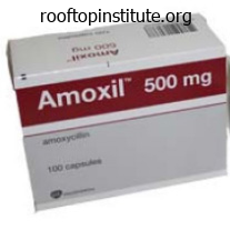
Amoxicillin 500 mg cheap fast delivery
In extra advanced lesions medications and side effects purchase amoxicillin 500 mg without prescription, the follicles are converted into cystic areas containing mucin treatment quincke edema purchase amoxicillin 500 mg free shipping, inflammatory cells, and altered keratinocytes. A perifollicular and intrafollicular infiltrate of lymphocytes, histiocytes and eosinophils is seen6. Although the existence of a primary form of follicular mucinosis has been questioned by some authors (who think about it as an "indolent" localized form of cutaneous T-cell lymphoma44), options in favor of a main form are the younger age of the affected person, a solitary plaque or limited number of lesions in the head and neck area, spontaneous resolution, and the absence histologically of epidermotropism and atypical lymphocytes. A wait-and-see strategy is really helpful for main follicular mucinosis as many circumstances resolve spontaneously inside 2�24 months. Treatment Mucinous Nevus A mucinous nevus (nevus mucinosus) is a benign hamartoma that might be congenital or acquired. Histologically, a diffuse deposit of mucin is seen in the higher dermis, and collagen and elastic fibers are absent inside the mucinous space. The dermis may be normal or it might be acanthotic with elongation of the rete ridges and hyperkeratosis, as in an epidermal nevus. The latter set of findings factors to a mixed hamartoma, by which features of an epidermal nevus are related to those of a connective nevus of the proteoglycan kind. Urticaria-like follicular mucinosis Urticaria-like follicular mucinosis is a really uncommon disorder that occurs primarily in middle-aged males. Pruritic urticarial papules or plaques seem on the top and neck within an erythematous "seborrheic" background. Hair-bearing regions could also be involved, however neither follicular plugging nor alopecia is seen. Urticaria-like follicular mucinosis waxes and wanes irregularly over a interval that may differ from a quantity of months to 15 years. Response to pure sunlight has been inconsistent, however it has been useful in a small variety of circumstances. As in major follicular mucinosis, mucin-filled cystic spaces occupy hair follicles. In the higher dermis, lymphocytes and eosinophils are seen round blood vessels and hair follicles. In only a single patient to date have been vascular C3 deposits seen by direct immunofluorescence. The prognosis is nice, and primarily based upon a restricted number of case stories, antimalarials and dapsone were reportedly beneficial46. It favors the trunk, head and neck, and genital area; hardly ever, cutaneous myxomas have an acral location48. When a number of, they could be a manifestation of Carney complex (cutaneous myxomas, cardiac myxomas, quite a few lentigines, multiple blue nevi, endocrine overactivity). Histologically, a cutaneous myxoma is a lobulated, well-defined lesion characterised by a mucinous matrix inside the dermis and the subcutis, with variably formed fibroblasts, mast cells, and some collagen and reticulin fibers. This differentiation is necessary as (angio)myxomas are true neoplasms, although benign, which can recur after incomplete excision. However, whatever the source, pathogenetic mechanisms, or underlying illness, amyloid materials shares certain widespread tinctorial and physico-chemical properties. He believed that the substance resembled starch or cellulose because, like starch, it turned blue when stained with iodine followed by dilute sulfuric acid. In 1928, Gutmann first described a patient with scientific features of lichen amyloidosis, while Freudenthal, in 1930, launched the term "lichen amyloidosus"three. Since 1980, there has been a marked decline within the incidence of symptomatic rheumatoid arthritis-associated secondary systemic amyloidosis as a end result of better management of the inflammatory response5,6. Primary cutaneous amyloidosis is usually observed in Southeast Asian international locations, together with Singapore, Taiwan, and Thailand7. Macular amyloidosis can also be commonly seen in Central and South American international locations, particularly those near the equator8. In the localized types, amyloid deposition occurs at or near the site of synthesis, whereas in the systemic varieties the precursors are secreted into the circulation, followed by amyloid deposition at distant sites9. Amyloidosis can also be categorized according to its constituent precursor protein (Table forty seven. Pathogenesis the most important element of amyloid is the fibril protein; the minor elements are amyloid P element, glycosaminoglycans, and apoE lipoprotein. Each condition is related to a selected precursor protein10; these amyloid precursors are initially soluble proteins that bear adjustments resulting in aggregation, polymerization, fibril formation, and, lastly, extracellular tissue deposition as insoluble amyloid. Deposits of amyloid may be seen in a range of medical problems, from plasma cell dyscrasias and Alzheimer disease to familial polyneuropathies and primary cutaneous lichen amyloidosis. In main cutaneous amyloidosis, the deposits are derived from keratin (macular, lichen, biphasic) or immunoglobulin gentle chains (nodular). The specific cutaneous lesions of major systemic amyloidosis are waxy, translucent or purpuric papules, nodules and plaques. Primary systemic amyloidosis is due to a plasma cell dyscrasia while secondary systemic amyloidosis arises from chronic inflammatory conditions such as rheumatoid arthritis or within the setting of continual infections. Prolonged friction, genetic predisposition, Epstein� Barr virus, and environmental elements have all been proposed as attainable etiologic components. This has been supported by ultrastructural research demonstrating transitional types between viable keratinocytes and amyloid, in addition to by constructive reactions with monoclonal antibodies directed towards basal layer keratins. An various theory suggests that the fabric is produced at the epidermo-dermal interface, with precursor proteins being secreted by basal keratinocytes. Although these cutaneous amyloid deposits stain positively with anti-human antibodies directed in opposition to IgG, IgM and IgA, this staining is assumed to be the outcomes of nonspecific immunoglobulin absorption as opposed to an immunoglobulin being the putative precursor protein. Apolipoprotein E4, galectin-7, and actin have also been proven to be related to major cutaneous amyloid deposits and could additionally be synthesized regionally by epidermal keratinocytes12,13. Small fiber neuropathy has been noticed in main cutaneous amyloidosis and may be associated to the related pruritus13a. Thus, its origin is very totally different from that of either macular or lichen amyloidosis11. Amyloid properties the method by which this transformation takes place differs amongst the various forms of amyloidoses. In major systemic amyloidosis, substitution of amino acids at particular positions inside the variable region of the immunoglobulin mild chain doubtlessly destabilizes these chains thereby rising the probability of their conversion to amyloid fibrils. Similarly, mutations in the transthyretin gene have been shown to considerably alter the stability of the transthyretin protein and enhance its baseline mild amyloidogenicity2,4. The accumulation of these relatively inert amyloid fibrils inside vital organs leads to strain atrophy and functional impairment. The commonest variants are macular amyloidosis, lichen amyloidosis, and biphasic amyloidosis. Detection of deposits inside clinically normal pores and skin permits analysis previous to medical symptoms (cerebral hemorrhage). More generally, heavy chains produce a non-amyloid sort deposit termed heavy chain deposition disease which is sometimes associated with acquired cutis laxa. Clinical features Although major cutaneous amyloidosis is classically divided into macular (macular amyloidosis), papular (lichen amyloidosis), and nodular (nodular amyloidosis) varieties, the first two entities actually represent the ends of a clinical spectrum. In some sufferers and even some lesions each macular and papular types could be present and this is termed "biphasic amyloidosis"15,16. Primary cutaneous amyloidosis can have a profound effect on quality of life due to the related pruritus and visible appearance of the pores and skin lesions17.
Syndromes
- Encephalitis
- A nasogastric (NG) tube thru the nose into the stomach to empty the stomach (gastric lavage)
- BUN
- Flank pain or back pain
- Drugs that affect platelet function
- Medications
- Anti-inflammatory medication (indomethacin)
Buy discount amoxicillin 250 mg on-line
No specific microorganism was incriminated in these circumstances and therapeutic trials have been empirical symptoms 8-10 dpo amoxicillin 250 mg generic with mastercard. A clinical trial of those relatively benign therapies could additionally be undertaken symptoms to pregnancy generic amoxicillin 500 mg overnight delivery, however no methodology of predicting a response has been recognized. The pure course of the illness is characterized by persistence for a quantity of years with eventual spontaneous remission in many sufferers. Although a remission rate of 10�15% was initially described24,forty two, further research have reported remission rates of 30�60%. There should be repeated makes an attempt to taper systemic medicines, on the prospect that a spontaneous remission has occurred. The web site within which every of these proteins resides determines the ultrastructural location where the blisters come up (Table 32. Its current name, "epidermolysis bullosa hereditaria", was coined by Koebner in 1886. The analysis is based on the medical phenotype, ultrastructural and immunohistochemical findings, and molecular genotype. Advances in our data of the pathogenesis of this group of diseases may lead to the development of simpler gene-, protein- and cell-based therapies. In laryngo-onycho-cutaneous syndrome, the underlying mutations affect solely the a isoform of the laminin three subunit. Although the resultant protein is structurally irregular, immunohistochemical staining of the dermal�epidermal junction is normally indistinguishable from that of regular skin. Extensive blisteringonthe buttocks(A)and blisteringinassociation withfocalkeratoderma onthesoles(B)ofa 2-year-oldgirl. Repeated blistering of the external eye can lead to neovascularization and blindness33. Chronic involvement of the esophagus results in scarring, stricture formation and, not often, even complete obstruction34. Involvement of the small intestine presents with continual malabsorption, whereas disease exercise throughout the giant intestine tends to produce constipation and anal fissures or strictures. Recurrent genitourinary tract blistering could result in urethral or ureterovesical strictures; if persistent, the latter might eventuate in ureteric reflux and hydronephrosis. If untreated, affected individuals lose enamel throughout childhood as a result of extreme caries38. Initially presenting as proximal internet formation between adjoining digits, the digits may ultimately turn into completely fused and encased by scar tissue. Lack of mobility results in bone resorption and muscular atrophy, and hand operate is severely compromised. Although the muscle illness presents during infancy in some sufferers, weakness usually develops insidiously throughout later childhood or even early maturity in those who are less severely affected. Renal illness could outcome from untreated outflow obstruction, glomerulonephritis, secondary systemic amyloidosis or IgA nephropathy. Although as but unproven, selenium or carnitine deficiency could additionally be a contributing factor. Although this can be a comparatively rare complication, careful surveillance throughout childhood is indicated. In addition, the severity, distribution and sort of pores and skin findings in a person affected person may change over time, such as evolution from generalized to localized blistering (or the opposite) or delayed growth of sequelae corresponding to exuberant granulation tissue, scarring, and nail dystrophy2. There is a restricted differential prognosis for a persistent mechanobullous illness with early onset and/or a positive family history2. The differential diagnosis of blisters and erosions in neonates or younger infants can also embody infectious ailments. Diagnostic concerns in sufferers with acral blistering, erosions, and ulcerations with progression to digital resorption may embrace congenital erythropoietic porphyria (see Table 32. For example, there are stories of the successful transfer of genes, corresponding to people who encode one of the chains of laminin 33249, into keratinocytes from affected individuals; when these genetically corrected keratinocytes are transplanted onto immunodeficient mice, the resultant epithelium has no evidence of blistering. Other investigators are finding out various sources, similar to mesenchymal stromal cells and inducible pluripotent stem cells, for the cellular correction of this disease56. Protective bandaging, padding over bony prominences, and soft/loosefitting clothes may be useful. Antibiotics should be used judiciously, with avoidance of continual therapy with topical mupirocin or oral antibiotics. Soft silicone dressings, some of which incorporate an absorbent foam backing, are extensively used. Silver-impregnated dressings may be useful for closely colonized or infected wounds, although long-term software should be averted to reduce systemic silver absorption. Less costly Vaseline-impregnated gauze is one other appropriate dressing for noninfected wounds. Tissue culture-derived artificial skin bioequivalents are additionally out there to be used in the management of continual, recalcitrant ulcers60. History In 1954, Theresa Kindler described a patient with acral blistering and photosensitivity throughout infancy, adopted later in life by progressive poikiloderma and atrophy. Twenty years later, Weary reported ten members of a single household with similar findings in addition to widespread atopic dermatitis throughout early childhood and keratotic papules over the joints of the arms and legs that appeared in childhood and continued indefinitely. Oral involvement and different mucosal manifestations were subsequently emphasized69�73. Pathogenesis Kindler syndrome is characterised ultrastructurally by basement membrane reduplication and combined planes of cleavage that can be within basal keratinocytes, by way of the lamina lucida, and/or under the lamina densa2,seventy four. Through integrin-mediated signaling, kindlin-1 affects the shape, polarity, adhesion, proliferation, and motility of keratinocytes74. There can also be proof that kindlin-1 has a role in regulating cutaneous epithelial stem cell homeostasis, with lack of kindlin-1 leading to increased risk of pores and skin cancer as well as cutaneous atrophy because of stem cell exhaustion and untimely senescence of keratinocytes77. ClinicalFeatures Erosions are occasionally present at start, most frequently on the forearms and shins, and blistering throughout infancy is most outstanding on the arms and feet. Reticulated hyperpigmentation and telangiectasias start to develop in sun-exposed areas throughout childhood, and after puberty these findings spread to sun-protected sites. Eczematous dermatitis, typically starting during infancy and resolving by early childhood, happens in some sufferers. Mucosal involvement might lead to intraoral and corneal scarring, ectropion, colitis, and strictures of the esophagus, urethra, vagina and anus. Pathology In older sufferers, pores and skin biopsy specimens present the everyday modifications of poikiloderma, including epidermal atrophy, vacuolization of the basal cell layer, variable epidermal melanin content, dermal melanophages, and capillary dilation. Immunostaining of pores and skin biopsy specimens with anti-kindlin-1 antibodies typically demonstrates a marked reduction or absence of this protein74. As poikiloderma turns into obvious throughout early childhood, the differential diagnosis may include Rothmund�Thomson syndrome and different entities outlined in Table 63. Mascar� Jr have discovered a decreased threshold for inducing suction blisters in these patients3,four.
Amoxicillin 500 mg order with mastercard
History Nikolsky first acknowledged the attribute histopathology of the bullous form of congenital ichthyosis in 1897 treatment math definition amoxicillin 500 mg order free shipping, and Brocq differentiated between dry (non-bullous) and wet (bullous) forms of congenital ichthyosiform erythroderma in 19022 symptoms flu amoxicillin 500 mg discount with amex. Based on the distinctive histopathologic features of the epidermis, Frost and Van Scott3 introduced the name "epidermolytic hyperkeratosis" for this autosomal dominant blistering form of congenital ichthyosis in 1966. It is usually an autosomal dominant disorder with full penetrance, with uncommon reports of autosomal recessive inheritance. A gentle perivascular lymphohistiocytic infiltrate is normally present in the higher dermis. Keratins 1 and 10 are coexpressed within the differentiated spinous and granular layers of the dermis, which are the sites of illness pathology on this disorder. Mutations in other sites are uncommon and customarily associated with a milder or unusual phenotype14. Mutations perturb keratin alignment, oligomerization, and filament meeting; this weakens the cytoskeleton, compromises the mechanical strength and cellular integrity of the dermis, and results in cytolysis and blistering. Epidermal acanthosis and hyperkeratosis outcome from hyperproliferation, decreased desquamation, and other factors. The barrier perform of the pores and skin is markedly disturbed, leading to elevated transepidermal water loss and bacterial colonization of the stratum corneum. Prenatal analysis may be performed when the underlying mutation has been identified in affected members of the family. Clinical distinction from the various forms of epidermolysis bullosa, staphylococcal scalded pores and skin syndrome, and other vesiculobullous and erosive issues that may current in neonates may require skin biopsy specimens and cultures (see Chs 32 & 34). However, sufferers may still periodically shed large plates of superficial dermis, revealing a young, erythematous base. In the neonatal interval, infants require administration in 897 Ichthyoses, Erythrokeratodermas, and Related Disorders larger deletions encompassing contiguous genes. The medical presentation varies considerably among sufferers and households, and six characteristic scientific patterns with or with out palmoplantar involvement have been described16. Sepsis as well as fluid and electrolyte imbalances may be life-threatening within the neonatal period. Episodes of blistering and secondary skin infections are additionally problematic later in life. Angular cheilitis and extreme scalp involvement resulting in encasement of hair shafts and hair loss are additional associated findings. Striking ortho-hyperkeratosis, intracellular vacuolizationof keratinocytes,anda prominentgranular layerwithclumped keratinfilamentsare 898 an intensive care nursery to present protective isolation and prevent or treat dehydration, electrolyte imbalance, and cutaneous superinfection. When the neonate is dealt with rigorously and protective padding and lubricants are used, erosions and denuded pores and skin usually heal rapidly. In youngsters and adults, remedy is aimed toward decreasing hyperkeratosis, removing scale, and softening the skin. Keratolytic lotions and lotions containing urea, salicylic acid, and -hydroxy acids are effective however often not well tolerated, particularly in kids, because of burning and stinging. Widespread topical software of higher-concentration salicylic acid preparations must be avoided because of the chance of systemic salicylism. Topical tretinoin and vitamin D preparations could additionally be helpful but typically trigger pores and skin irritation. Bacterial skin infections are common and infrequently set off blistering; they require topical or systemic antibiotic remedy. Use of antiseptics corresponding to antibacterial soaps, chlorhexidine, or dilute sodium hypochlorite baths might assist to control bacterial colonization. Continuous use of oral or topical antibiotics must be prevented because of the risk of developing antibiotic resistance. Low initial doses with gradual enhance and cautious monitoring are advisable, with use of the bottom effective maintenance dose18. Trauma-induced small blisters on the extremities happen during infancy however often subside by early childhood, while hyperkeratosis develops. A attribute function is superficially denuded areas with collarette-like borders, described as "molting" or "Mauserung", which develop because of superficial blistering and shedding of the stratum corneum. Pathology Histopathologic abnormalities embody orthokeratotic hyperkeratosis, acanthosis, and vacuolization of the granular cell layer that sometimes results in superficial intraepidermal separation. By electron microscopy, clumping of tonofilaments is current however limited to the upper spinous and granular cell layers. Histologic findings in ichthyotic skin embody acanthosis, perinuclear vacuolization of keratinocytes within the higher epidermis, and parakeratotic hyperkeratosis. The ensuing mutant keratin 10 protein has an arginine-rich carboxy terminus, which redirects the protein from its normal cytoplasmic location to the nucleolus. Each island of normal pores and skin represents a revertant clone that arises via mitotic recombination, i. The actual position of the frameshift mutations in the markedly elevated fee of mitotic recombination remains to be determined. Pathology Most histopathologic options, including orthokeratotic hyperkeratosis, hypergranulosis, acanthosis, and church spire-like papillomatosis, are nonspecific. Since the initial description, a quantity of large households with autosomal dominant inheritance and sporadic circumstances have been reported. Because the latter lacks the same old glycine loop motifs, protein�protein interactions are altered23. A well-known instance of the ichthyosis hystrix phenotype is the "porcupine males" of the Lambert household from Suffolk, England26. Clinical expression varies even inside families, starting from isolated palmoplantar keratoderma (which could also be severe and mutilating) to localized hyperkeratotic plaques to generalized hystrix-like hyperkeratosis with attractive, inflexible spikes. Circular constriction bands (pseudoainhum), starfishlike keratoses, knuckle pads, digital flexion contractures, and secondary bacterial infections have been described. Collodion infants who finally develop lamellar ichthyosis have been shown to harbor mutations within the transglutaminase-1 gene, leading to deficiency of this significant cross-linking enzyme of the epidermis27. Transglutaminase-1 deficiency perturbs formation of the cornified cell envelope, leading to hyperkeratosis, profoundly impaired barrier perform, and transepidermal water loss. Treatment Collodion infants are at risk for thermoinstability, hypernatremic dehydration, pores and skin infections, and sepsis. It is therefore important to maintain Clinical Features Collodion babies are sometimes born prematurely and have increased perinatal morbidity and mortality. Its tautness often results in ectropion, eclabium, and hypoplasia of nasal and auricular cartilage. Sucking and pulmonary air flow may be impaired, leading to dehydration, malnutrition, hypoxia, and pneumonia. In the method, fissures develop that impair epidermal barrier perform, which might result in percutaneous lack of water and permit the entry of microorganisms; fluid and electrolyte imbalances, skin infections, and sepsis could comply with. In addition, round bands of hardened skin can result in vascular constriction and distal edema. Within two to four weeks, the membrane peels off in sheets and a transition to the underlying disease phenotype takes place.
Generic amoxicillin 250 mg otc
Lymphomatoid drug reactions develop insidiously over a interval of months or even years after initial administration of the culprit drug symptoms breast cancer amoxicillin 250 mg buy amex. Cutaneous lesions could additionally be solitary or multiple medications during pregnancy 250 mg amoxicillin buy with mastercard, localized or generalized, and include erythematous to violet papules, plaques, or nodules. Numerous widespread tumors are rare as is an erythroderma simulating S�zary syndrome. Histologically, a dense lymphocytic infiltrate is seen within the dermis that can mimic a T- or B-cell lymphoma. In some patients, the lymphocytic infiltrate is band-like, resembling mycosis fungoides. Atypical nuclei with a cerebriform outline and epidermotropism may be observed. In the lymph nodes, focal necrosis and eosinophilic and histiocytic infiltrates might destroy the conventional architecture. Lesions resolve inside weeks to months following withdrawal of the accountable treatment. A B melanin or iron and in some situations, higher described as discoloration; and (3) simply postinflammatory changes. Exposure to heavy metals such as silver and gold in addition to arsenic may also induce darkening of the pores and skin, and bleomycin can lead to linear "flagellate" hyperpigmentation. Hypopigmentation can happen with the continual use of several topical medicines, together with retinoic acid and corticosteroids; depigmentation is related primarily with the application of monobenzyl ether of hydroquinone and imiquimod or exposure to catechols, phenols, and quinones. Cutaneous hypopigmentation can result from oral tyrosine kinase inhibitors, in particular imatinib and cabozantinib. For example, chloroquine, imatinib, dasatinib and sunitinib can lead to lightening and even depigmentation (see Table 21. Drug-induced psoriasis Drugs may be associated with the precipitation or exacerbation of psoriasis. A drug can have an effect on the affected person with psoriasis in a quantity of ways: (1) exacerbation of pre-existing psoriasis; (2) induction of lesions of psoriasis in clinically regular skin in a person with psoriasis; (3) precipitation of psoriasis de novo; and (4) growth of therapy resistance59. The medical manifestations of drug-induced psoriasis span the spectrum of psoriasis, from limited or generalized plaques to erythroderma and pustulosis of the palms and soles. A big selection of drugs have been implicated in the induction or exacerbation of psoriasis. Lesions of drug-induced psoriasis normally regress inside weeks to a number of months of discontinuing the inciting drug. The latter happen not just in people with psoriasis or rheumatoid arthritis, but also in sufferers being treated for other circumstances. Additional unusual drug reactions Examples of such drug reactions are outlined in Table 21. Cutaneous Side Effects of Vaccines and Injected Medications Vaccine-induced reactions With the discontinuation of vaccinations for smallpox in the general inhabitants, the incidence of serious cutaneous unwanted facet effects due to vaccines is now low (see Ch. Lichenoid eruptions, erythema multiforme, and infrequently autoimmune reactions. In addition, the generally administered influenza vaccine has been associated with a serum sickness-like reaction, acute febrile neutrophilic dermatosis, and linear IgA bullous dermatosis. Occasionally, native abscess formation may follow vaccination of strongly reactive individuals, administration of a considerable amount of vaccine, or a deep injection. A which permits identification of the offending agent with certainty, the choice is often made to discontinue all medication which are non-essential in addition to the "high-probability" medication. For mild drug eruptions, topical corticosteroids and antihistamines could additionally be useful. Supportive interventions include warming of the environment, correction of electrolyte disturbances, high caloric supplementation, and prevention of sepsis. The presumed immunologic etiology for drug eruptions has led to using systemic corticosteroids, immunosuppressives, and anticytokine therapies. Finally, after recovery, patients ought to be advised to avoid the drug thought to be responsible for the response and all chemically associated compounds. The variety of lesions and their distribution pattern should also be assessed, together with whether or not all the lesions are purpuric or simply these located on the distal lower extremities. This article serves as an introduction to a way for analysis and classification of patients presenting with purpura, outlined as visible hemorrhage into the skin or mucous membranes. The differential diagnosis presented on this chapter is directed towards syndromes of primary purpura, the place the hemorrhage is an integral a half of lesion formation, somewhat than secondary hemorrhage into established lesions. Lesions within the first three groups are categorized on the basis of size, while those in the final three teams can vary in measurement from a quantity of millimeters to a quantity of centimeters in diameter. Two main causes of purpura, microvascular occlusion syndromes and vasculitis, are discussed in Chapters 23 and 24. The former are essential to recognize because they may mimic vasculitis however require a really completely different strategy to prognosis and therapy. The most typical presentation of microvascular occlusion syndromes is non-inflammatory retiform purpura (see Table 22. Early lesions seldom show much erythema, and in the uncommon occasion by which early erythema is present, purpura or necrosis usually includes a minimal of two-thirds of the lesion. In the skin, livedo reticularis is a mirrored image of the physiologic anatomy of gradual move states (see Ch. It is the three-dimensional construction and move regulation of the dermal and subcutaneous vasculature that offers rise to the net-like pattern of livedo reticularis. The diameter of the almost-circular individual units within the net-like grid varies from 2 cm or larger on the again to 5 mm or less on the palms or soles. Retiform purpura morphology results from occlusion of the vessels that produce the livedo reticularis pattern, but the two entities may be distinguished based mostly upon the presence or absence of purpura, respectively, therefore the time period "retiform purpura"1. Given the dimensions of dermal vessels, the clot within the vessel is often too small to be seen grossly. What is actually observed is hemorrhage around the vessel throughout the dermis, presumably due to ischemia with hemorrhage prior to full occlusion of the vessel. The shape of such a hemorrhagic lesion is determined by the anatomy of the slow flow network, though an entire reticulate pattern may be very hardly ever seen. Instead, the morphology of retiform purpura is composed of "puzzle pieces" of the livedo reticularis sample. This strategy to the differential prognosis of purpura represents a departure from traditional categorization by pathophysiology. Because the pathophysiology of purpuric syndromes is what the clinician is making an attempt to confirm, sorting by pathophysiology is of limited clinical utility. A methodology based primarily on the morphology of purpuric lesions (in addition to quantity and distribution) is designed to streamline the process of producing medical hypotheses and more than likely diagnoses, thereby facilitating a rapid, environment friendly and accurate evaluation to show or disprove the suspected diagnosis. The Time Course of Purpura the three subsets of macular (non-palpable) purpura (see Tables 22. As such, these lesions have a quite simple evolution, from preliminary hemorrhage to steady clearing of red blood cells and hemoglobin. Clinically, this correlates with fading of lesions and, in larger lesions, transition of colour from red�blue or purple to green, yellow or brown earlier than complete fading.
Buy amoxicillin 500 mg visa
Vasculitis of medium-sized blood vessels is characterized by similar adjustments involving the vessels medicine lodge treaty amoxicillin 250 mg discount free shipping. Neovascularization of the adventitia medicine disposal buy discount amoxicillin 500 mg online, in the type of small capillaries, is usually seen in older lesions of medium-sized vessel vasculitis. Immunoglobulin deposition is highest (up to 100%) in skin lesions current for 48 hours4. However, there are additional types of cutaneous vasculitis, in particular lymphocytic (Table 24. Use of the term lymphocytic vasculitis often requires clarification, especially in discussions with non-dermatologists. Sneddon syndrome Lymphocytic thrombophilic arteritis (macular arteritis) Panniculitis, together with lupus Table 24. Rhizopus) Lichenoid capillaritis (pigmented purpura) Cellulitis Hemorrhage � Trauma � Solar (actinic) purpura * � Medication-related. Lesions often begin as a purpuric macule or partially blanching urticarial papule. Residual postinflammatory hyperpigmentation may persist for months after the first course of resolves. In general, signs or signs of gastrointestinal, renal, or neurologic involvement ought to enhance the medical suspicion for a systemic vasculitis. In one examine, the presence of paresthesias or fever and the absence of painful lesions were recognized as risk components for an related systemic disease11. In the latter group, the average duration of disease exercise was 24�28 months3,four. The presence of arthralgias or cryoglobulinemia and an absence of fever might portend chronicity11. The need for and choice of further therapies depends on the severity of the cutaneous involvement, chronicity, and whether or not systemic involvement is current. Oral dapsone (50�200 mg/day) can lead to improvement of gentle to average chronic lesions. Patients with extreme, ulcerating, or progressive cutaneous illness who require fast management of signs could be treated with a brief course of high-dose oral corticosteroids. Because of the multiple opposed results of long-term oral corticosteroids, a taper over 4 to 6 weeks ought to be tried. The traditional tetrad consists of palpable purpura, arthritis, stomach ache, and hematuria. The common age of onset is 6 years, and 90% of instances happen in kids <10 years of age14,15. The disorder follows a seasonal pattern, with a peak prevalence in the course of the winter. Lack of glycosylation of the hinge area of IgA1 could promote the formation of macromolecular complexes that lodge within the mesangium and activate the alternate complement pathway14. Urticaria, vesicles, bullae, targetoid lesions, and foci of necrosis may also be seen. Typically, lesions are symmetrically distributed on the buttocks and decrease extremities, however may also involve the trunk, higher extremities, and face. Individual lesions often regress inside 10 to 14 days, with resolution of cutaneous involvement over a interval of several weeks to months. Gastrointestinal involvement (50�75% of patients) could precede the purpura and presents with colicky belly pain (65%), gastrointestinal bleeding (30%), and/or vomiting. Renal involvement happens in 40�50% of sufferers and typically presents with microscopic hematuria (40%), usually accompanied by proteinuria (25%). Although the appearance of cutaneous lesions usually precedes the event of nephritis, the latter is clinically evident inside 3 months14. In pediatric sufferers, threat factors for the event of nephritis embrace age >8 years at onset, abdominal pain, and recurrent disease19. Depending upon the collection, persistent renal disease has been noticed in 8�50% of patients16, emphasizing the need for longitudinal monitoring until all abnormalities resolve. The lung is also a uncommon web site of involvement, presenting as hemoptysis and/or pulmonary infiltrates due to diffuse alveolar hemorrhage20. For example, necrotic pores and skin lesions are present in 60% of adults whereas cutaneous necrosis is noticed in <5% of children21,22. There are conflicting information as to whether or not the chance of renal insufficiency is elevated if purpura are current above the waist21,23. In contrast, 60�90% of adult sufferers with neoplasm-associated IgA vasculitis will have cancer of a solid organ, in particular the lung25�27. Adults are also more doubtless than children to have diarrhea and leukocytosis, to require extra aggressive remedy, and to have a longer hospital stay28. In one sequence of adults with IgA vasculitis (age >40 years), an absence of eosinophils almost tripled the risk of renal involvement29. Perivascular C3 deposits and papillary dermal edema had been associated with renal involvement in a single sequence of pediatric patients29a. Referral to a nephrologist is acceptable for sufferers with evidence of renal involvement. In one research, using prednisone was related to a extra rapid resolution of renal illness during the 4-week therapy period34. This evaluation additionally found no distinction in the danger of persistent renal disease in patients with severe kidney illness who have been treated with cyclophosphamide versus supportive care. Lastly, a meta-analysis that examined using systemic corticosteroids on the time of diagnosis versus supportive care found a decrease in the mean (but not the median) time to decision of belly ache, whereas the odds of creating persistent renal illness were decreased39a. Cutaneous lesions usually start as plaques with variable levels of hemorrhage and favor the top and extremities. The typical interval between the inciting event (see above) and the onset of illness is 1�2 weeks43. Cutaneous involvement presents abruptly with massive erythematous patches or urticarial plaques that then evolve into medallion, annular, iris, or targetoid purpuric plaques. Arcuate, polycyclic, scalloped, or rosette-shaped lesions and vesicobullae happen much less generally, as does therapeutic with atrophic scars43. Tender, nonpitting edema of the face, ears, extremities (including the palms and feet), and scrotum is attribute. Although mucosal and visceral involvement is uncommon, oral petechiae, conjunctival injection, abdominal ache, arthralgias, glomerulonephritis, and intussusception (<1%) may occur40. The course is benign, with spontaneous and complete resolution with out sequelae within 1 to three weeks. Acute hemorrhagic edema of infancy � triggers Infections � Adenovirus � Coxsackievirus � Cytomegalovirus � Epstein�Barr virus � Herpes simplex virus � Hepatitis A virus � Measles � Rotavirus � Varicella zoster virus � Campylobacter spp. In young kids with urticarial lesions and acrofacial edema, urticaria multiforme ought to be considered44. The differential prognosis also includes erythema multiforme, urticaria, urticarial drug eruptions, serum sickness-like reactions, Kawasaki disease, urticarial vasculitis, and Sweet syndrome. Given the numerous cutaneous hemorrhage, trauma, acute meningococcemia, and purpura fulminans are typically thought-about. Routine laboratory tests are nonspecific, and the prognosis is based on acceptable clinicopathologic correlation.
Common Centaury (Centaury). Amoxicillin.
- How does Centaury work?
- Dosing considerations for Centaury.
- What is Centaury?
- Are there safety concerns?
- Loss of appetite and stomach discomfort.
Source: http://www.rxlist.com/script/main/art.asp?articlekey=96411
Discount 500 mg amoxicillin
This dysfunction is brought on by mutations within the gene encoding loricrin symptoms 5 dpo amoxicillin 250 mg with visa, a glycine-rich cornified envelope protein33�35 (see Ch medications for ocd amoxicillin 500 mg free shipping. Generalized desquamation or features of a collodion child may be evident at delivery, with subsequent evolution to a mild generalized ichthyosis. However, the absence of deafness as well as the presence of ichthyosis distinguish loricrin keratoderma from Vohwinkel syndrome39. The differential analysis can also embrace ichthyosis hystrix Curth�Macklin as a outcome of mutations within the variable tail area of keratin 1 (see Ch. Histologic options of loricrin keratoderma embody hyperkeratosis with parakeratotic cells, scattered transitional cells, and a broad stratum granulosum with focal perinuclear vacuolization38,39. The ensuing proteasome insufficiency disturbs terminal epidermal differentiation and interferes with processing of profilaggrin. Other options embrace flexion deformity of the fingers with associated sclerosis and parallel linear arrays of keratotic papules in the flexural areas of the extremities. Histologic analysis reveals orthohyperkeratosis, focal parakeratosis, acanthosis, and hypergranulosis with attribute irregular keratohyalin granules. Immunostaining for filaggrin is positive in the cornified layer quite than the granular layer. Management Stronger keratolytic remedy may be useful, similar to 5�10% salicylic acid in white delicate paraffin, or 5�6% salicylic acid in 70% propylene glycol gel; occlusion for a couple of nights per week enhances efficacy, and mechanical debridement can be carried out. Responses to oral erythromycin and topical tacrolimus have been described in the Bothnia and Nagashima types, respectively30,31. Histologic options of the keratoderma embody orthohyperkeratosis, acanthosis, and hypergranulosis. A characteristic finding is sort of full absence of Langerhans cells within the affected pores and skin. Other manifestations embody sclerodactyly with variable cutaneous atrophy resembling Huriez syndrome67, nail dystrophy. Connexin 26 is expressed in the palmoplantar epidermis, sweat glands, and cochlea of the internal ear. Patients have nonprogressive high-tone sensorineural listening to loss, which may be missed throughout infancy53. The dysfunctional connexin 30 disturbs cell�cell communication in epithelial cells. Although first reported in a large FrenchCanadian kindred, Clouston syndrome has also been described in a selection of other ethnic groups. Small papules in a grid-like array that corresponds to eccrine acrosyringia could also be seen, especially on the distal digits where the dermatoglyphs are most outstanding. A, Courtesy, Alfons Histologic findings are nonspecific, with papillomatosis and prominent orthohyperkeratosis. Progressive hypotrichosis that may eventuate in full alopecia affects the scalp, eyebrows, eyelashes, and axillary and genital areas; the hair is often wiry, brittle, and pale. The nail abnormalities could mimic these of pachyonychia congenita or other syndromes of "hair�nail hypoplasia"seventy one. Histologic analysis of thickened palms and soles exhibits orthohyperkeratosis with a normal granular layer. Papular lesions may reveal proliferation of ductal constructions within a fibrovascular stroma. Ultrastructural examination of the hair shows disorganization of the hair fibers, with a loss of the hair shaft cuticle. Erythema and warty hyperkeratosis develop in the perioral and perianal areas as properly as in other intertriginous sites80, leading to pain and pruritus81. Keratoses in linear streaks and with a perifollicular distribution also appear in the flexures of the extremities82,83. Histology of affected pores and skin demonstrates acanthosis with orthohyperkeratosis and parakeratosis, papillomatosis, and a perivascular inflammatory infiltrate; an elevated variety of mast cells has additionally been observed86. Pseudoainhum and hyperkeratotic psoriasiform plaques on the elbows and knees often develop. An further feature is periodontitis with onset during childhood, which leads to untimely loss of teeth. There is a predisposition to pyogenic infections of the skin and internal organs, together with hepatic abscesses92. Haim�Munk syndrome is an allelic situation that also options onychogryphosis, arachnodactyly, and acro-osteolysis93,94. Oral retinoid remedy not only lessens the hyperkeratosis but additionally decreases the risk of periodontal and infectious complications98,ninety nine. Patients with Sch�pf� Schulz�Passarge syndrome additionally develop hidrocystomas of the eyelids and different adnexal tumors. The zinc metalloprotease encoded by this gene is crucial for cholesterol homeostasis and 934 this autosomal recessive disorder described in families from the Greek island of Naxos is defined by keratoderma, woolly hair, and a lifethreatening cardiomyopathy100,101. A member of the armadillo protein household, plakoglobin hyperlinks the inside and outer parts of the desmosomal plaque by binding to the cytoplasmic domain of cadherins. The right ventricular cardiomyopathy has just about 100 percent penetrance, typically manifesting with arrhythmias, heart failure, and/or sudden demise throughout adolescence. Lesions often develop in adolescence or early adulthood and are exacerbated by mechanical stress. There is usually an excellent response to oral acitretin and/or software of keratolytic agents112. Wedge-shaped subungual hyperkeratosis leads to elevation, thickening, and darkening of the nail plate, which may assume an "omega" form with extra outstanding involvement distally than proximally. There may or is most likely not involvement of all of the digits, however the toes, thumbs, and index fingers are inclined to have extra severely affected nails. In infants, erythema of the nail mattress can precede the looks of dystrophic modifications. Thick yellow plantar keratoses develop in sites of strain, with the formation of calluses, fissures, and blisters in areas of friction, notably in the course of the summer season; spread to the dorsal ft occasionally occurs in association with trauma or an infection. Severe pain typically makes it difficult for sufferers to walk, and palmoplantar hyperhidrosis and paronychia are frequent findings. Laryngeal involvement can result in hoarseness and occasionally respiratory tract obstruction125. Pathology In hyperkeratotic pores and skin and oral plaques, the keratinocytes in the superficial epidermis and mucosal epithelium have a attribute pale cytoplasm and eosinophilic inclusions. At the ultrastructural level, these findings correlate with perinuclear condensation of mutated keratin filaments and vacuolization of the peripheral cytoplasm124. Similar histologic findings are seen in white sponge nevus, which is brought on by keratin 4 and 13 mutations.
Purchase amoxicillin 250 mg without prescription
It begins during the first few days to weeks of life within the setting of transient myeloproliferative disorder treatment 2 degree burns 250 mg amoxicillin purchase with visa, a congenital leukemoid response that affects roughly 10% of neonates with Down syndrome symptoms 10 weeks pregnant 250 mg amoxicillin cheap mastercard. Vesicles and pustules are located most prominently on the face but can also contain the trunk and extremities29. Histologic features of the vesicular and papulopustular lesions embody intraepidermal vesicles containing eosinophils, perivascular infiltrates of eosinophils, and an eosinophilic folliculitis similar to that seen in eosinophilic pustular folliculitis19. Histologically, intraepidermal spongiotic vesiculopustules are current in affiliation with a perivascular infiltrate. The infiltrate includes immature myeloid cells, which can be evident in a Tzanck smear of vesiculopustules and are normally additionally current in the peripheral blood29,30. Although the skin lesions are most likely to resolve spontaneously inside weeks to months as the leukemoid response subsides, affected patients are at increased danger for the event of myeloid leukemia through the first few years of life and thus should be followed by a pediatric oncologist31. Infants with congenital erosive and vesicular dermatosis must be adopted for proof of diminished sweating and neurologic defects. Treatment is supportive with meticulous wound care, as no specific remedy is on the market. Pyoderma Gangrenosum Rarely reported in infants and newborns, pyoderma gangrenosum in this age group most frequently involves the perineum, possibly because of pathergy. Otherwise, the scientific features of the ulcer, together with an undermined violaceous border, are similar to those seen in older kids and adults35,36. Biopsy specimens present a neutrophilic infiltrate with out evidence of infection, but the diagnosis is often considered one of exclusion after other causes of ulceration such as an infection, immunodeficiency, thrombosis, and vasculitis are eliminated. Tense bullaonthewristand flaccidbullaeonthe dorsalhandaswellas pinkplaquesonthe trunkina1-month-old infant(seeTable34. Restrictive Dermopathy Restrictive dermopathy is a uncommon disease characterized by rigid, tense pores and skin with erosions and linear tears that happen most commonly in flexural creases. Other options embody translucent skin with outstanding vessels, micrognathia, sparse/absent eyelashes, natal tooth, large fontanelles, dysplasia of the clavicles, and poorly expanded lungs on account of the restrictive skin modifications. Most affected newborns die from restrictive pulmonary disease throughout the first week of life. The second type of noma has been seen in Native American infants who present with perineal and oral ulcerations in the setting of extreme combined immunodeficiency42. The presence of oral and perineal ulcerations in newborns and infants ought to prompt an analysis for an infection and immunodeficiency. Perinatal Gangrene of the Buttock this uncommon situation is characterized by the sudden onset of erythema and cyanosis of the buttocks, followed by progressive improvement of gangrene and ulceration. Although some instances have been related to umbilical artery catheter infusions, others were spontaneous. Occlusion or spasm of the inner iliac artery has been postulated as a potential cause43. A affected person with a unilateral congenital buttock ulceration related to dimples on the knees has additionally been reported44. Considerations when evaluating a neonate with hemorrhagic bullae and gangrenous soft tissue infarction also embrace purpura fulminans because of congenital coagulopathies. The first is a manifestation of an infection, usually Pseudomonas aeruginosa septicemia, and has been reported nearly exclusively in low-income countries40. Infants current with an abrupt onset of gangrenous ulcerations of the nose, lips, mouth, eyelids, anus, and scrotum. In severe circumstances, the ulcerations can destroy underlying bony buildings and trigger in depth deformity. Predisposing factors embrace untimely birth, low start weight, malnutrition, and previous illness. Some authors counsel that this form of noma neonatorum could characterize a variant of neonatal ecthyma gangrenosum41. Pustules, hyperpigmented macules,and collarettesof scaleareseenon thechinand neckofan African-American neonate. These apoeccrine sweat glands possessed sure morphologic and functional features of eccrine and apocrine glands. However, latest research that examined serial histologic sections from management and hyperhidrosis patients, using routine and immunofluorescent staining, discovered no evidence of apoeccrine glands1,2. The eccrine secretory unit consists of a proximal coiled secretory portion in the decrease dermis and subcutaneous tissue. Both cell varieties are surrounded by myoepithelial cells, which in all probability function to improve the delivery of sweat to the pores and skin floor. The ductal epithelium consists of two or more layers of cuboidal cells with out surrounding myoepithelium. The intraepidermal portion of the duct, the acrosyringium, is twisted like a corkscrew with related coils in the stratum corneum4. Innervation of the eccrine glands is supplied by postganglionic sympathetic fibers which have acetylcholine (not norepinephrine) as their principal terminal neurotransmitter (Table 35. The sweat middle responds to its own temperature (as a reflection of core physique temperature) as properly as neural stimuli from the periphery. Disorders of sweating are frequent and can be due to dysfunction of central sweat centers; sympathetic ganglia and their pre- and postganglionic fibers; or secretory glands and ducts. Hyperhidrosis, which can be emotional or secondary to systemic illness, often results in social stigmatization. Knowledge of the construction and performance of sweat glands is essential for proper analysis and remedy of sweating issues (see Chs 39 & 159). Functional eccrine glands are present at start and react to thermal and emotional stimuli. Function Eccrine sweat is a sterile, dilute electrolyte resolution that accommodates primarily sodium chloride (NaCl), potassium and bicarbonate. In addition, different organic compounds and heavy metals such as arsenic, cadmium, lead, and mercury are excreted in sweat7a. Keratinocyte outgrowths from eccrine sweat glands also have a role in re-epithelialization of human wounds9. In addition to their well-established function in thermoregulation, eccrine sweat glands have immunomodulatory, antimicrobial and excretory capabilities. Apocrine sweat is an odorless viscous fluid that incorporates precursors of odoriferous substances. Sebaceous glands contribute to skin barrier function, produce androgens, and have antimicrobial in addition to pro- and anti inflammatory activities. Apoeccrine glands are thought to have the same innervation and receptor profile as eccrine glands. Pilosebaceous unit with an associated apocrine sweat gland and a nearby eccrine sweat gland. The amount and quality of eccrine sweat secretion varies greatly, depending on emotional and environmental stimuli. Sweat is shaped in two steps10: (1) release of nearly isotonic major sweat by the secretory coil; and (2) partial reabsorption of NaCl by duct cells, ensuing within the delivery of a hypotonic fluid to the pores and skin surface. The ultimate focus of NaCl may be greater when sweat is produced at a extra fast rate.
Discount amoxicillin 250 mg without prescription
Hopf actually described nail changes and palmoplantar keratotic papules in his preliminary report of acrokeratosis verruciformis43 medicine joint pain amoxicillin 250 mg buy otc, and there have been subsequent observations of families the place some members had solely acrokeratosis verruciformis whereas others exhibited the classic skin findings of Darier disease46 treatment 001 amoxicillin 500 mg purchase. Because sufferers with Darier disease often develop warty lesions on the extremities earlier than keratotic papules seem in a seborrheic distribution, some people who current with acrokeratosis verruciformis may later be recognized as actually having Darier disease7. P602L, A698V) have been identified in sufferers with isolated acrokeratosis verruciformis as properly as those with acral warty lesions adopted by the onset of Darier illness after intense solar exposure12,47. Acantholysis is as a outcome of of a disturbance in cell adhesion that results in suprabasilar cleft formation. At the ultrastructural level, this corresponds to a loss of desmosomes and detachment of keratin filaments from the desmosomes41. Dyskeratosis is as a result of of apoptosis of keratinocytes and is characterized by nuclear condensation and perinuclear keratin clumping. Two kinds of dyskeratotic cells are observed in Darier disease42: "Corps ronds" � acantholytic enlarged keratinocytes in the malpighian layer with darkly staining and partially fragmented nuclei surrounded by a transparent cytoplasm and encircled by a brilliant ring of collapsed keratin bundles. Improvement following treatment with topical calcineurin inhibitors has also been reported54. Intermittent use of topical antibiotics and antifungal agents is helpful in reducing malodor because of microbial colonization and mild secondary infections. Several case reports have instructed that topical 5-fluorouracil (1% or 5%) may be efficacious55. However, the utilization of these agents is limited by their unwanted facet effects, and relapse happens after cessation of remedy. Because of the teratogenic potential of systemic retinoids, contraception is mandatory for ladies of childbearing potential (see Ch. Individual dose titration is necessary to optimize the benefit�risk balance and avoid excessive dose-related unwanted effects, similar to mucocutaneous dryness and elevation of serum lipids and liver enzymes. Intermittent remedy to stop exacerbations in the course of the summer season months is a helpful strategy. Oral contraceptives might mitigate symptoms in women with premenstrual exacerbations60. A family historical past may also be a clue to the prognosis of Darier illness, and histologic findings differentiate it from seborrheic dermatitis. However, in a persistent form of acantholytic dermatosis that impacts patients with vital actinic injury, the papules are scalier and the histologic features carefully resemble those of Darier disease48. The presence of palmoplantar papules, longitudinal erythronychia, distal notching of the nails, mucosal lesions, and outstanding dyskeratosis in addition to acantholysis histologically can function clues to the prognosis of Darier illness. Occasionally, a sporadic dysfunction referred to as "papular acantholytic dyskeratosis" may be localized to the vulvocrural area. Surgical remedy Surgical remedy could additionally be an efficient alternative for focal recalcitrant lesions, significantly in the flexural and gluteal areas. Destructive remedy must include the follicular infundibulum to be able to prevent recurrences. Experience is necessary to keep away from scarring, notably in physique areas in danger for hypertrophic scar or keloid formation. In small case collection, enchancment has been observed following pulsed dye laser and photodynamic therapy64. Daily skincare includes the utilization of antimicrobial cleansers to stop malodorous bacterial colonization and keratolytic emollients to reduce scaling and irritation. Topical remedy 950 Topical retinoids are more practical as monotherapy49,50 than topical vitamin D analogues51, which had no benefit in a small randomized managed study52, or corticosteroids. However, retinoid-associated irritation is common and may be decreased by alternate-day application, History In 1939, Howard and Hugh Hailey, brothers working within the Department of Dermatology at Emory University School of Medicine in Atlanta, Georgia, described a chronic dermatosis in two sets of brothers. The dysfunction was characterized by recurrent blisters and erosive, crusted lesions on the neck (in one pair of brothers) and within the axillae and groin (in the second pair)65. Histologic sections from all 4 sufferers confirmed similar options � intraepidermal vesicles, mild dyskeratosis, hyperkeratosis, and a reasonable dermal lymphocytic infiltrate. Different dermatopathologists interpreted these findings variably as pemphigus, pemphigus-like, or Darier disease. The authors themselves rightly believed that they had been dealing with a novel entity unrelated to Darier disease and coined the name "familial benign continual pemphigus". A illness known as "pemphigus cong�nital familiale h�r�ditaire" had been described 6 years previous to the report by the Hailey brothers. The subsequent controversy as to whether or not "familial benign continual pemphigus" was a novel illness or a phenotypic variant of Darier illness was eventually solved by our understanding of the molecular genetics of both problems. The mode of inheritance is autosomal dominant with full penetrance, however the age of onset and expressivity varies markedly amongst affected family members67,sixty eight. Particular mutations may be associated with primarily genitoperineal involvement69 or milder phenotypes70. Clinical Features Onset and scientific pattern the initial lesions and associated signs normally develop through the second or third decade of life however may be delayed till the fourth or fifth decade67. The scalp, antecubital and popliteal fossae, and trunk are much less regularly involved81. Development of persistent, moist, malodorous vegetations and painful fissures is widespread. Longitudinal leukonychia of the fingernails could serve as a refined diagnostic clue in patients with limited or atypical disease83. Symptoms Painful intertriginous erosions and crusting may interfere with actions of every day residing. Modifying components Given the abnormal cell�cell adhesion within the dermis, friction can induce new lesions. Microbial colonization and secondary infections are important modifying elements, as mentioned under. Course of the disease Complete remissions as well as flares are frequent, and the scientific course in a person patient is troublesome to predict. Some sufferers report attenuation of the illness at an older age, whereas others expertise no change or worsening with aging81. Thus, larger areas of dyscohesion with single or teams of acantholytic cells are seen, which have been likened to a "dilapidated brick wall". In distinction to Darier disease, necrotic keratinocytes are unusual, and solely uncommon acantholytic dyskeratotic cells. In more persistent lesions, epidermal hyperplasia, parakeratosis, and focal crusts are discovered. A reasonable perivascular lymphocytic infiltrate is observed within the superficial dermis. Complications As in Darier disease, colonization and secondary bacterial, fungal, and viral infections play an necessary position in illness exacerbation and persistence89,ninety. Topical and/or systemic antimicrobial brokers could additionally be required to induce a clinical remission. It is characterised by fever and a quickly spreading vesicular eruption, and ought to be promptly treated with an oral antiviral medication. Lesions confined to the axillae or groin could additionally be mistaken for intertrigo or candidiasis.
500 mg amoxicillin discount with mastercard
The first accounts of occupational pores and skin ailments targeted on unwell well being among miners medications used to treat depression 250 mg amoxicillin generic with amex, and in 1556 symptoms zollinger ellison syndrome amoxicillin 500 mg cheap without prescription, Georg Agricola detailed the deep ulcers observed amongst metal workers. Eleven years later, Paracelsus discussed the etiology, pathogenesis and remedy of skin changes attributable to salt compounds. In 1700, Bernardino Ramazzini printed a detailed treatise on the ailments of tradespeople by which he described pores and skin disorders incurred by tub attendants, bakers, gilders, midwives, millers, and miners6. Physicians began to extra widely acknowledge occupational irritant dermatitis in the course of the Industrial Revolution with the event of new materials and chemical compounds, each natural and synthetic, for both industrial and household use7. Germany and France have been the first to enact legal guidelines compensating staff for industrial pores and skin ailments. Although skin illness accounted for only 17% of all recorded non-fatal occupational diseases in 200712,13, it still ranks second and is only surpassed by musculoskeletal issues. The annual incidence of occupational contact dermatitis reported by dermatologists was zero. In addition to manufacturing, industries with the best nonadjusted and adjusted rates of dermatitis embrace healthcare and social assistance; arts, leisure and recreation; and lodging and food providers. Rubber chemical substances, soaps and cleansers, moist work, resins, acrylics, and nickel are a few of the more common sources for irritant and allergic contact dermatitis. These include concentration, pH, mechanical strain, temperature, humidity, and length of contact. Overall, the impact of occupational pores and skin illnesses on each society and the individual is formidable in mild of each prevalence and financial components. Determination of the inciting irritant or irritants is crucial to the successful diagnosis and subsequent therapy of this frustrating, and at times disabling, dysfunction. The protean vary of scientific shows varies from sensory disturbances with out visible skin findings to severe chemical burns and ulcers. The right analysis rests on an evaluation of crucial objective indicators of disease coupled with an insightful targeted history. Preventative measures embody work course of change, personal protective tools, and maximizing the barrier defenses of the dermis. In addition, irritated pores and skin could turn into more vulnerable to superimposed allergic sensitization. Important predisposing characteristics of the individual include age, sex, pre-existing skin illness, anatomic region exposed, and sebaceous activity. Lastly, essentially the most commonly affected websites are uncovered areas such because the palms and the face, with hand involvement seen in ~80% of patients and facial involvement in 10%14. Acute reactions involve direct cytotoxic damage to keratinocytes, while repeated exposures to solvents and surfactants cause slower harm to cell membranes by removing surface lipids and water-retaining substances. Cold alone may reduce the plasticity of the horny layer, with consequent cracking of the stratum corneum. It is characterized by one or more of the following indicators: scaling, redness, vesicles, pustules and erosions, usually beginning underneath occlusive jewelry. In this fashion, as quickly as activation is initiated via epidermal cells, continuous T-cell activation impartial of the exogenous agent may be maintained and barrier dysfunction perpetuated22. Damage to the stratum corneum lipids (which mediate barrier function) is associated with lack of cohesion of corneocytes, desquamation, and an increase in transepidermal water loss. Transepidermal water loss is among the triggering stimuli that promote lipid synthesis, keratinocyte proliferation, and transient hyperkeratosis in the course of the restoration of the cutaneous barrier. However, damage with a solvent can disrupt this protecting mechanism by occlusion and blockage of water evaporation, thus halting lipid synthesis and barrier recovery. Clinical symptoms develop solely after the cumulative damage exceeds an individually decided manifestation or elicitation threshold, which can decrease with progression of the disease. However, if the identical irritant exposures comply with each other carefully in time, or when the manifestation threshold is lowered. Signs may include xerosis, erythema and vesicles, however lichenification and hyperkeratosis predominate. Elderly individuals who regularly bathe without remoisturizing are at specific risk of growing asteatotic dermatitis. Intense pruritus is widespread, with the pores and skin appearing dry with ichthyosiform scale and characteristic patches of superficially cracked skin (see Ch. Patients must be asked whether they have cleansed the pores and skin with strong soaps or detergents. It is characterized by eczematous lesions, mostly on the arms, that final for weeks to months with persistent redness, infiltration, scale, and fissuring within the affected areas. The latter is able to profound reactions on the pores and skin floor: oxidation, discount, desiccant, vesicant, or ion disruption26. The acute response reaches its peak rapidly, often inside minutes to hours after publicity, after which begins to heal. These lesions are restricted to the world the place the irritant or toxicant damaged the tissue, with sharply demarcated borders and asymmetry pointing to an exogenous cause. This syndrome ought to be thought of in circumstances in which folliculitis or acneiform lesions develop in settings outdoors of typical pimples, particularly in patients with atopic dermatitis, seborrheic dermatitis, or prior zits vulgaris. Miliarial reactions, which can turn into pustular, can develop in response to occlusive clothing, adhesive tape, or ultraviolet and infrared radiation. Irritants capable of eliciting this response embrace propylene glycol, hydroxy acids, ethanol, and topical drugs such as lactic acid, azelaic acid, benzoic acid, benzoyl peroxide, mequinol, and tretinoin. This response to irritants such as lactic or sorbic acid may be reliably reproduced with dose responsiveness in double-blinded exposure exams. The frictional response contains hyperkeratosis, acanthosis, and lichenification, usually progressing to hardening, thickening, and elevated toughness. ContactUrticaria Contact urticaria is divided into non-immunologic and immunologic subtypes (see Ch. Irritants that may produce immunologic contact urticaria include parabens (preservatives), henna, ammonium persulfate (oxidizing agent), and latex. Non-immunologic contact urticaria may be caused by a selection of exposures, from caterpillars and jellyfish to stinging nettles and meals (see Table 15. Risk elements embody atopy, hand dermatitis, previous mucosal exposure to latex. Over time, extra histologic findings embody acanthosis with gentle hypergranulosis and hyperkeratosis. Principally, all strong acids give the same medical options, together with erythema, vesication, and necrosis. Inorganic acids are generally utilized in business, especially hydrofluoric, sulfuric, hydrochloric, chromic, nitric, and phosphoric acids (Table 15. Hydrofluoric acid and sulfuric acid trigger essentially the most severe burns, even at low concentrations, and there may be important absorption leading to systemic toxicity30.
Buy 500 mg amoxicillin
Mucositis of the oral and gastrointestinal mucosae is a significant medicine syringe buy amoxicillin 250 mg visa, usually dose-limiting symptoms 39 weeks pregnant amoxicillin 250 mg purchase visa, poisonous impact of chemotherapy and it might be notably severe when chemotherapy is combined with radiotherapy. Candida, herpes simplex virus) and/or hemorrhage, reflections of bone marrow suppression. Topical corticosteroids, bodily barriers, decreased sun publicity, and broad-spectrum sunscreens may show helpful. Preventive measures include sustaining oral hygiene via using soft toothbrushes and rinsing with water and sodium bicarbonate. In addition to particular antimicrobial brokers, topical anesthetics could additionally be helpful; in some patients, systemic analgesics are required. Occasionally, palifermin is prescribed, however its use can result in white coating of the tongue and transient keratoses. The frequency of extravasations has been markedly decreased by the use of "permanent" central venous catheters, whereas the incidence of acral erythema has increased in the setting of prolonged infusions of drugs and the continual use of oral multi-kinase inhibitors (see below). A variety of phrases have been used to describe the cutaneous poisonous effects of chemotherapy. Long-term administration of hydroxyurea is related to the development of painful ulcerations of the malleolar area of the lower extremities. Targeted remedy using focused therapies that act on cell-surface receptors and intracellular signaling pathways is rapidly increasing55. The unwanted effects of targeted therapies and newer antineoplastic agents are outlined in T ables 21. Immunotherapy As with targeted agents, using immunotherapies is increasing exponentially. They are being used for common malignancies similar to nonsmall-cell lung cancer in addition to metastatic melanoma. Drug-Induced Adverse Reactions Involving Hair and Mucosae Hair A appreciable variety of medicines induce hair loss53. They affect the hair follicles via two major mechanisms: anagen effluvium (abrupt interruption of the energetic progress phase) and telogen effluvium (an increased variety of hairs within the resting phase). The hair loss is often reversible after discontinuation of the responsible agent. Diagnosis of drug-induced telogen effluvium could be difficult, and requires statement of improvement after the suspected drug has been withdrawn. With regard to drug-induced hair progress, it could be very important distinguish between hypertrichosis and hirsutism. Mucosal ulcerations Stomatitis with erosions and ulcerations may occur both as a half of a drug-induced mucocutaneous syndrome. Ulcerative stomatitis has also been observed with metamizole, phenylbutazone, bisphosphonates, oral calcineurin inhibitors, D-penicillamine, gold salts, and the vasodilator nicorandil56. In addition, allergic reactions to dental materials and publicity to metals corresponding to mercuric chloride or copper sulfate might trigger stomatitis. Also referred to as papulopustular eruption; could be related to secondary Staphylococcus aureus or Demodex infection/infestation. Contact irritancy from urine containing medicine corresponding to foscarnet can lead to penile ulceration. Other Drug-Induced Cutaneous Reactions Acneiform eruptions (including folliculitis) Acneiform eruptions represent ~1% of drug-induced pores and skin eruptions. Less commonly, azathioprine, quinidine, and adrenocorticotropic hormone are the culprits. Anticoagulant-induced skin necrosis Anticoagulant-induced skin necrosis is a uncommon, doubtlessly life-threatening reaction induced by warfarin or heparin. Warfarin-induced necrosis usually begins 2 to 5 days after remedy is initiated and coincides with the anticipated early drop in protein C function (see Ch. One in every 10 000 individuals who receives warfarin will develop this facet impact, with middle-aged obese women and those with a hereditary deficiency of protein C being at highest threat. Clinically, erythematous painful plaques evolve into hemorrhagic blisters and necrotic ulcers as a consequence of ischemic infarcts. The latter are as a result of occlusive thrombi inside blood vessels of the skin and subcutaneous tissue2. The most typical websites of involvement are the breasts, thighs, stomach, and buttocks. Therapy includes discontinuing warfarin and administering vitamin K, heparin (as the anticoagulant), and intravenous infusions of protein C focus. Heparin-induced cutaneous necrosis is due to antibodies that bind to complexes of heparin and platelet issue four and induce platelet aggregation and consumption (see Ch. Platelet counts are often depressed, but, until the baseline platelet depend is thought, this is probably not appreciated. The interval between drug publicity and the acneiform eruption depends on the offending agent. Discontinuation of the heparin and administration of anticoagulants, similar to argatroban or danaparoid, is beneficial. Granulomatous reactions Granulomatous drug reactions can mimic interstitial granulomatous dermatitis and granuloma annulare and are discussed in Chapter ninety three. These opposed cutaneous reactions, especially when extreme, necessitate discontinuation of the drug. In syndromes of inflammatory hemorrhage, such as cutaneous small vessel vasculitis, in addition to in microvascular occlusion, the evolution and clearing of lesions is more complicated. Conversely, a late lesion of occlusion with some resulting dermal necrosis could on histologic examination show features characteristic of leukocytoclastic vasculitis. The scientific and histologic assessments of purpuric lesions are equally important in properly evaluating and diagnosing a purpuric syndrome. When a platelet plug is insufficient because of the size of the vessel or the dimensions of the damage, secondary hemostasis with clot formation is required. The management of clot formation is critically important: too little clotting can result in dying by hemorrhage; inappropriate clotting produces thrombosis, embolus or necrosis; and uncontrolled clotting with fibrinolysis can produce each thrombosis and hemorrhage, as in disseminated intravascular coagulation. The number, distribution, and morphology of purpuric lesions are necessary elements in producing a probable and efficiently testable scientific hypothesis. Basal coagulation encompasses the constant low-level activation of some parts of the procoagulant, natural anticoagulant, and fibrinolytic pathways, so that these methods are prepared for a rapid response when wanted. This section might take place on any cell that expresses tissue issue and is exposed to plasma; inflammatory cells and injured endothelial cells are common sites. Only small quantities of these factors are released on this phase, normally insufficient to produce a clot. However, these small quantities are important for triggering the propagation/amplification part of coagulation. In addition, fibrinolytic exercise is generated through both activation of single- and two-chain urokinase-like plasminogen activator and launch of tissue plasminogen activator, which subsequently convert plasminogen to plasmin. C1 esterase inhibitor, well known in controlling complement reactions, also has a significant function in regulating this pathway. Of note, the episodic edema of hereditary angioedema is probably due more to poor C1 esterase inhibitormediated down-regulation of bradykinin manufacturing than to deficient inhibition of complement (see Ch. The contact activation system is likely responsible for a few of the fibrinolysis and refractory hypotension seen in sepsis, following cardiopulmonary bypass ("post-pump" systemic inflammatory response syndromes), and in other settings the place contact activation is prone to be intensive.
